
the Creative Commons Attribution 4.0 License.
the Creative Commons Attribution 4.0 License.
 | 08 Oct 2026
Gut microbiomes of the little free-tailed bat (Mops pumilus) show high prevalence of potential bacterial pathogens but limited responses to land cover
Julie Teresa Shapiro

Christopher Barnes
Ida Broman Nielsen
Linett Rasmussen
Ara Monadjem
Robert A. McCleery
Anders J. Hansen
Land cover change is a threat to wildlife globally. It may also impact the gut microbiome harbored by wildlife, with implications for their health, conservation, and One Health more broadly. Bats are a diverse group of mammals that can be found in a range of habitats, with differing degrees of anthropogenic disturbance. Here, we analyzed the V3–V4 region of 16S rRNA from fecal samples from 109 individual little free-tail bats (Mops pumilus) in northeastern Eswatini. We aimed to describe the species composition, richness, and community structure of the gut microbiome and identify and potentially pathogenic bacteria that M. pumilus might harbor. We then measured the effects of host characteristics, land cover composition and configuration on the species richness, phylogenetic diversity, prevalence of potentially pathogenic genera, and community composition of the gut microbiome. We found high levels of individual variability and no significant differences in species richness, phylogenetic diversity, or community composition based on sex, age, or reproductive condition. We detected five potentially pathogenic bacterial genera, four at high prevalence: Mycoplasma (88 %), Rickettsia (36 %), Salmonella (34 %), Bartonella (23 %), and Campylobacter (4 %). The co-occurrence of two to five of these genera within an individual was common. Using a subsample of 83 individuals captured at 17 distinct roosts, we found a small influence of fine-scale land cover on the community composition but none on the richness or phylogenetic diversity of the gut microbiome. We did not find associations between land cover composition, configuration, or host characteristics with the prevalence of potentially pathogenic genera. Our results demonstrate the complex influences on bat gut microbiome, while the high prevalence of potential pathogens shows the importance of continued research and surveillance.
- Article
(1096 KB) - Full-text XML
-
Supplement
(1335 KB) - BibTeX
- EndNote


Humans have drastically changed the face of the Earth, and one of the primary ways is through land cover change (Feddema et al., 2005; Leemans and Zuidema, 1995; Song et al., 2018), the conversion of Earth's surface (Wulder et al., 2018). Land cover change threatens biodiversity, contributes to climate change, and may increase the emergence of infectious diseases (Daszak et al., 2001; Lambin et al., 2010; Leemans and Zuidema, 1995). These changes have profound impacts on wildlife through the reduction of available habitats and resources (Fischer and Lindenmayer, 2006; Tscharntke et al., 2012), edge effects, patch isolation, and loss of connectivity across the landscape (Fahrig, 2003).
Land cover change affects not only wildlife populations but also individuals and the microbiome they harbor. The microbiome consists of microorganisms, including bacteria, Archaea, protozoa, and fungi that live on or in organisms (Leonard et al., 2024; Ley et al., 2008b; Zilber-Rosenberg and Rosenberg, 2008). This is essential for an organism's health as it may affect metabolism, nutrients acquisition, energy uptake and storage, immune system function, susceptibility to pathogens, aging, and longevity (Bäckhed et al., 2004; Biagi et al., 2016; Claesson et al., 2012; Ley et al., 2006; Turnbaugh et al., 2007). The gut microbiome in turn is shaped by numerous factors, including host phylogeny (Ley et al., 2008a, b; Sanders et al., 2014), genetics (Goodrich et al., 2014), diet (Carmody et al., 2015; Ley et al., 2008b; Muegge et al., 2011; Storm et al., 2024), social interactions (Raulo et al., 2024), and the surrounding environment (Bernardo-Cravo et al., 2020; Rothschild et al., 2018).
In a range of wildlife species, land cover change may reduce individuals' gut microbiome diversity or alter its composition, likely due to modified diets (Barelli et al., 2015; Chang et al., 2016; Ingala et al., 2019; Mistrick et al., 2024), altered environmental microbiomes (Bernardo-Cravo et al., 2020), and direct or indirect interactions with domestic animals (Fackelmann et al., 2021). There is evidence in some wildlife species that reduced gut microbiome diversity and altered community structure can have negative effects on animals' fitness (Suzuki, 2017; Worsley et al., 2021) and their immune systems (Amato et al., 2013; Siddiqui et al., 2022), leaving them vulnerable to disease (Jiménez and Sommer, 2017; Warne et al., 2019) or parasites (Schmid et al., 2022; Weldon et al., 2015). In fact, this relationship between gut microbiome and disease is characterized by complex interactions and feedback loops as a healthy gut microbiome provides a first line of defense against colonization by pathogens, prevents opportunistic pathogens that may be present from multiplying, and contributes to healthy immune system function (Bernardo-Cravo et al., 2020). Dysbiosis, often characterized by altered community composition and shifted beta diversity, leaves hosts vulnerable to infections, which may then further alter the gut microbiome, increasing the risk of co-infections and repeated infections with various types of pathogen (Schmid et al., 2022; Weldon et al., 2015). However, defining dysbiosis in wildlife remains challenging, if not impossible, for most species (Handy et al., 2023). Observed patterns, such as reduced alpha diversity or shifts to beta diversity, are often interpreted as indicative of potential dysbiosis, although explicit signs of resulting pathology or dysfunction are often not evident or measured. Nevertheless, it is likely that all of these factors have cumulative effects on wildlife populations and their conservation. Thus, a better understanding of how land cover change affects the gut microbiome of wildlife is important for conservation, wildlife health, and One Health (Muhummed et al., 2025; Trevelline et al., 2019) – the principle that the health of the environment, animals, and humans is interconnected (Mettenleiter et al., 2023).
Bats are the second-most diverse order of mammals, with 1500 described species (Burgin et al., 2025; Simmons and Cirranello, 2025). They are threatened globally by land cover change (Frick et al., 2020; Pekin and Pijanowski, 2012), yet relatively little is known about how this affects their microbiome. In the common vampire bat (Desmodus rotundus), land cover change is associated with a shift in diet from wildlife to livestock (Ingala et al., 2019). While the subsequent effects on alpha diversity and richness in this species' microbiome appear marginal (Fleischer et al., 2024), land cover change may lead to shifts in the core microbiota, microbiome heterogeneity (Ingala et al., 2019), and abundance of specific bacterial genera (Fleischer et al., 2024). In Glossophaga soricina, a Neotropical frugivore, individuals feeding in banana monoculture in Costa Rica showed less individual variation and had less diverse microbiomes than those foraging in organic banana farms and forests, which the authors interpreted as potential indicators of dysbiosis (Alpízar et al., 2021). For Pipistrellus kuhlii in Catalonia, Spain, urban and agricultural land covers were associated with lower alpha diversity (Lobato-Bailón et al., 2023).
These changes in the bat microbiome may also have important implications from a One Health perspective. Many bat species are known to host many different viruses (Ge et al., 2012; Geldenhuys et al., 2018; Hayman, 2016; Shapiro et al., 2021; Wu et al., 2016), which could be pathogenic to humans, other animal species, or the bats themselves. While studies are currently limited, there is some evidence that enteric viral infections may be linked to decreased gut microbial diversity and altered community composition. Further, such viral infections may cause dysbiosis in the gut and leave bats vulnerable to co-infections with other pathogens (Melville et al., 2025; Wasimuddin et al., 2018) or from potentially pathogenic bacteria found in the gut microbiome itself (Szentivanyi et al., 2023), with negative effects such as reduced body condition found in some cases (Melville et al., 2025). Bartonella, Leptospira, Mycoplasma, Rickettsia, Anaplasma, Borrelia, and Coxiella are the most commonly identified potential pathogens, although their pathogenicity and capacity to spill over to other species are often unknown (Szentivanyi et al., 2023). These bacteria may cause fatal disease in bats (Evans et al., 2009; Imnadze et al., 2020), potentially threatening populations. Further, Myotis species in northern Europe (Veikkolainen et al., 2014) and Pteropus species in New Caledonia (Descloux et al., 2021) have been identified as reservoirs of Bartonella mayotimonensis and Mycoplasma haemohominis, respectively, both of which cause disease in humans.
The savannas of southern Africa are home to a high diversity of bat species (Monadjem et al., 2020; Monadjem et al., 2021) but are severely threatened by land cover change, particularly conversion to agriculture, including both lower intensity small-holder crops and intensive commercial monocultures (Aleman et al., 2016; Laurance et al., 2014). This land cover change can decrease bat diversity and activity, although the effects vary widely between foraging guilds and species (Mtsetfwa et al., 2018; Shapiro et al., 2020; Swartz et al., 2022; Weier et al., 2018, 2021), with open-air foragers in particular using and even preferring these habitats (Mtsetfwa et al., 2018; Noer et al., 2012; Shapiro et al., 2020). While the year-round availability of water and insect prey in monocultures may provide apparently favorable habitat for these bats (Shapiro et al., 2020; Swartz et al., 2022; Weier et al., 2018), this could essentially be an ecological trap (Tanalgo et al., 2025) with potential changes in foraging altering their microbiome (Alpízar et al., 2021; Lobato-Bailón et al., 2023).
Thus, with its implications for maintaining healthy bat populations and preventing disease spillover, describing the bat gut microbiome and understanding how it is affected by land cover change is important. As a model, we use the little free-tailed bat Mops pumilus, a widespread species found in both natural and anthropogenic land covers (Monadjem et al., 2020) whose gut microbiome has not yet been characterized. We collected samples from 109 individuals in northeastern Eswatini (formerly Swaziland), a hotspot of bat diversity (Monadjem et al., 2021) undergoing rapid land cover change (Bailey et al., 2015; Shapiro et al., 2020). Our objectives were to (1) describe the species composition of the gut microbiome in M. pumilus; (2) identify potential bacterial pathogens and their prevalence; (3) determine the role of both host characteristics (age, sex, reproductive condition) and land cover metrics on gut microbiome richness, phylogenetic diversity, potential pathogen prevalence, and community composition. We expected anthropogenic land covers and fragmentation to decrease the species richness of the microbiome and alter the community composition (Amato et al., 2013; Barelli et al., 2015; Lobato-Bailón et al., 2023). We also expected to find a higher prevalence of potential pathogens in areas with higher proportions of anthropogenic land covers and fragmented savanna (Daszak et al., 2001; Lambin et al., 2010; Mistrick et al., 2024).
2.1 Study area
This study was conducted in the Lowveld region of eastern Eswatini, bordered by the Drakensberg Mountains in the west and the Lubombo Mountains in the east (Fig. 1). The area is a part of the Maputaland-Pondoland-Albany biodiversity hotspot (Monadjem et al., 2021; Steenkamp et al., 2005) with an elevation ranging from approximately 150 to 600 m above sea level. Land cover is dominated by commercial sugarcane plantations, subsistence maize fields, grazing lands for domestic livestock, rural villages, and several protected parks (Monadjem and Reside, 2008; Shapiro et al., 2020). It has been subject to rapid land cover change, particularly due to the expansion of commercial sugarcane plantations and small-holder agriculture (Bailey et al., 2015). The dominant vegetation type prior to agricultural transformation was open savanna or woodland (Monadjem et al., 2003).

Figure 1Map of the study area and land cover types. Sites of bat capture are indicated by the numbered points. Tick marks and values correspond to Universal Transversal Mercator (UTM) coordinates (zone 36S), equivalent to approximately 10 km.
To quantify variability in the land cover of our study site, we used Google Earth Engine (https://earthengine.google.com, last access: 27 November 2016) to classify the landscape based on using a Landsat 8 8 d raw composite image from 21 to 29 March 2016 at 30 m resolution. We classified the image using a voting support vector machine (voting SVM) classifier based on 193 training points for the following four categories: rural settlements (hereafter “rural”), savannas, sugarcane plantations (hereafter “sugarcane”), and water (Shapiro et al., 2020). To further distinguish savanna vegetation from crops and pasture in rural areas (Prestele et al., 2016), we overlaid the population count raster for Eswatini (Swaziland) from WorldPop projected for 2015 (Linard et al., 2012) and reclassified cells with population count >1 as rural (Shapiro et al., 2020).
2.2 Bat capture and sample collection
We collected bats under a permit from the Eswatini National Trust Commission. Handling methods were approved by the Institutional Animal Care and Use Committee (IACUC) at the University of Florida (Protocol #201508751).
We surveyed bats from November 2015 to July 2016 at a total of 28 different sites reflecting the variation of land cover in the area. Capture sites included 17 roosts and 11 other areas of likely bat activity not directly near a roost (trails, clearings, edges of lakes, reservoirs, streams, irrigation, etc.). To capture bats, we used mist nets (EcoTone) measuring 12 m×3 m and/or one harp trap at each site (Fig. 1). We identified bats to species according to distinctive physical characteristics and measurements as described in Monadjem et al. (2020). To aid in the identification of species, we measured the forearm length of each captured bat to the nearest 0.1 mm with calipers and recorded mass to the nearest 0.5 g with a spring balance. We recorded the sex of each bat. We assessed reproductive condition as follows: females were classified as pregnant, lactating (producing milk, enlarged nipples, no surrounding fur), post-lactating (enlarged nipples not producing milk, fur regrowth), or non-reproductive. Males were classified as reproductive (enlarged, visible testes) or non-reproductive (Racey, 1974). We determined age (juvenile or adult) by examining calcification of the epiphyseal joints (Kunz and Anthony, 1982; Racey, 1974).
We placed captured bats individually in cloth holding bags for the deposition of feces. All bags were soaked in a diluted bleach solution prior to the first use and between each use (Shapiro et al., 2024). Bats were released after the collection of feces. We then placed fecal samples in Eppendorf tubes with silica gel to desiccate them and preserve DNA. Samples remained in a freezer at −10 °C until the end of the field season in July 2016, after which we transferred them to a −20 °C freezer.
2.3 DNA extraction
We extracted DNA using the MoBio PowerViral Environmental RNA/DNA Isolation kit according to the manufacturer's instructions, with the following modifications: samples were vortexed in a Tissuelyzer II (Qiagen) at a frequency of 30 Hz for 10 min and subsequently centrifuged at 13 000×g for 2 min.
We ran an initial SYBR Green chemistry quantitative polymerase chain reaction (qPCR) on a subset of 10 samples and two blanks to optimize cycle number and ensure that inhibitors co-purified with the DNA would not bias the results (Bustin et al., 2009). The qPCR was done in a dilution series of extract (1 µL undiluted, 1 µL 1:2, 1 µL 1:5, 1 µL 1:10). We carried out qPCRs on an Agilent Technologies Stratagene Mx3005P quantitative PCR thermocycler using the Amplitaq Gold enzyme system. The 25 µL reactions consisted of 1 µL DNA, 0.2 µL Amplitaq Gold (5 U µL−1, ThermoFisher Scientific), 2.5 µL 10× Gold PCR Buffer (ThermoFisher Scientific) and 2.5 µL MgCl2 (25 mM, ThermoFisher Scientific), 0.2 mM dNTP Mix (25 mM, Invitrogen), 1 µL BSA (20 mg mL−1, New England Biolabs, Inc.), 1.5 µM each of forward and reverse 16S primer, and 1 µL of SYBR Green/ROX solution consisted of 1 part SYBR Green I nucleic acid gel stain (S7563) (Invitrogen), 4 parts ROX reference dye (12223-012) (Invitrogen) and 2000 parts high-grade DMSO.
We performed PCR on extracts with tagged bacterial 16S ribosomal RNA primers (16S) forward (341F): 5'-CCTAYGGGRBGCASCAG-3' and reverse (806R): 5-GGACTACNNGGGTATCTAAT-3; each primer had a unique 6 nucleotide base tag added to the 5-end (Hansen et al., 2012), and each tag combination was used only once through this study (e.g., no repetition of any tag combinations). These primer sets flank the V3–V4 region of 16S rRNA of bacteria, resulting in a PCR product of approximately 440 bp. We ran three separate PCR reactions for each sample.
We performed PCR reactions in a total volume of 25 µL with 3 µL DNA, 0.2 µL Amplitaq Gold (5 U µL−1, ThermoFisher Scientific), 2.5 µL 10× Gold PCR Buffer (ThermoFisher Scientific) and 2.5 µL MgCl2 (25 mM, ThermoFisher Scientific), 0.2 mM dNTP Mix (25 mM, Invitrogen), 1 µL BSA (20 mg mL−1, New England Biolabs, Inc.), 1.5 µM each of forward and reverse primer. We ran all samples for 25 cycles in Thermocyclers (Applied Biosystems 2720 Thermal Cycler) with the following settings: 95 °C – 5 min; (95 °C – 15 s; 55 °C – 30 s; 72 °C – 40 s) × 25 cycles; 72 °C – 4 min; 4 °C hold. We visualized all PCR products (5 µL) on 2 % agarose gel stained with GelRed Nucleic Acid Gel Stain (Biotium). We then carried out separation by electrophoresis with 140 V, 350 mA for 40 min to determine the presence of the desired target. We included negative controls of extraction and PCR blanks in all PCRs, electrophoresis, and sequencing reactions to rule out contamination.
We then pooled PCR products based on band intensity from the gel electrophoresis by adding 5 µL of samples with bright bands to the pool and 10 µL of samples with weaker bands. We made a total of nine pools, each containing 35–45 samples. We purified pooled samples with the QIAquick PCR Purification Kit (Qiagen) as preparation for library building. We built a library on each of the nine pools with the Illumina TruSeq DNA PCR-Free Library Preparation Kit according to the manufacturer's manual. We also made a negative library control. We purified libraries with Beckman Coulter Agencourt AMPure XP (1:1, library: bead ratio) to remove primer dimers. We ran the purified libraries on an Agilent Technologies 2100 Bioanalyzer using the Agilent High Sensitivity DNA kit to determine the length of the products and concentration for pooling of libraries for sequencing. We pooled libraries in equimolar ratios based on results from the Bioanalyzer. Pooled libraries were sequenced on an Illumina MiSeq 250 bp paired-end sequencer at the Danish National High-Throughput Sequencing Centre, Centre for GeoGenetics, Copenhagen, Denmark.
2.4 Bioinformatic processing
We demultiplexed the libraries to samples using a custom script (available from http://github.com/tobiasgf/lulu, last access: 17 March 2021) (Frøslev et al., 2017) that assigns reads to samples based on an exact match of both the forward and reverse tags. Adapters, primers, and internal tags were also removed during demultiplexing using CutAdapt (v1.9.1) (Martin, 2011). We removed unpaired reads below 100 bp.
Reads were demultiplexed using the DADA2 pipeline (Callahan et al., 2016) within the R environment (version 4.1.2) (R Core Team, 2013) following Barnes et al. (2020) using custom scripts (available from https://github.com/jtshapiro/Mops_pumilus_Gut_microbiomes, last access: 18 September 2026) (Shapiro, 2026). Demultiplexed reads were deposited in the Sequence Read Archive (SRA), the National Center for Biotechnology Information (NCBI; BioProject PRJNA1420193), and are freely available for download. The pipeline performs quality filtering by calculating error rates and removing reads above the threshold. We identified and removed chimeras using the removeBimeraDenovo function in DADA2. Finally, we assigned taxonomy to each amplicon sequence variant (ASV) using DADA2's native naïve RDP Bayesian classifier against the Silva 138 database (Quast et al., 2012).
We checked negative controls for the number of reads and their species content. Any ASVs identified in negative controls were removed from further analysis. We further filtered the ASV table to remove any non-bacterial ASVs (eukaryotes and Archaea). We counted ASVs as occurring in a sample only if it was detected in at least two out of three PCR replicates because those found in only a single replicate may be the result of sequencing errors or contamination (Ficetola et al., 2015; Willerslev et al., 2014).
In order to visualize the completeness of our survey, we calculated a sample-based rarefaction curve for all samples together using the function “specaccum” in the vegan package (Oksanen et al., 2007) (Fig. S1 in the Supplement). We then repeated this for each individual bat to ensure that our results and analyses regarding individual species richness were not biased by sequencing depth. As the species accumulation curves for both the full data set and all individual bats reached an asymptote (Fig. S2), we included all individual bats in downstream analyses without exclusions or rarefaction.
All downstream analyses were based on the presence/absence of ASVs and their relative abundances.
2.5 Statistical analysis
All statistical analyses were completed using custom scripts (available from https://github.com/jtshapiro/Mops_pumilus_Gut_microbiomes) (Shapiro, 2026) in R (version 4.1.2) (R Core Team, 2013).
2.5.1 Landscape metrics
We measured landscape metrics around all roost capture sites following Shapiro et al. (2020), using the “ClassStat” function in the SDMTools package (VanDerWal et al., 2014). To measure land cover composition, we calculated the proportion of land cover of each type (rural, savanna, sugarcane, and water). We also measured two landscape configuration metrics: savanna edge density, because many bats use edges of natural vegetation (Chambers et al., 2016; Ethier and Fahrig, 2011; Mendes et al., 2017; Müller et al., 2012); and savanna splitting index (hereafter “savanna splitting”), to account for the fragmentation of savanna vegetation, which has a positive effect on M. pumilus activity in this region (Shapiro et al., 2020).
We calculated these metrics at the fine scale (120 m buffer radius) and landscape scale (2 km buffer radius) as both scales are relevant to explaining the activity of M. pumilus in the region (Shapiro et al., 2020). The fine scale captures local conditions around the bats' roosts. The larger scales correspond to the scale at which M. pumilus responds to the environment, and its range of home ranges and nightly foraging distances (Noer et al., 2012; Shapiro et al., 2020). Only bats captured at roosts (n=83) were included in models using landscape metrics.
2.5.2 Bacterial species identity, richness, and phylogenetic diversity
We measured bacterial species richness for each individual bat. Species richness was measured by summing the total number of ASVs per bat. We counted the most common bacterial phyla and orders across all individual bats. We calculated estimated bacterial species richness of the full data, including all bats, set using the Chao1 Estimator (Chao, 1987) with a confidence interval of 95 % using the function “spp.est” in the fossil package (v.0.3.7) (Vavrek, 2011). We then calculated Faith's phylogenetic diversity (Faith, 1992) for each bat. To do so, we first aligned all the ASV sequences using the ClustalW method (Thompson et al., 1994) with the function “msa” in the MSA package (v.1.26) (Bodenhofer et al., 2015). Using the seqinr package (v.4.2-30) (Charif and Lobry, 2007), we converted the msa object to an alignment with the function “msaConvert” and then calculated the pairwise distance between all the aligned sequences with the function “dist.alignment”. Based on these distances, we built a neighbor-joining tree (Saitou and Nei, 1987) using the function “nj” in the ape package (v.5.7-1) (Paradis et al., 2004). Finally, we calculated the phylogenetic diversity within each bat using the function “pd” in the picante package (v.1.8.2) (Kembel et al., 2010).
We analyzed differences in both ASV richness and phylogenetic diversity between bats based on sex and age (adult or juvenile) using Welch's two sample t test, which is robust to unequal sample sizes and variances between groups (Ruxton, 2006; Welch, 1947). We then analyzed differences based on reproductive condition (non-reproductive females or males, lactating, post-lactating, or pregnant females, scrotal males), which may influence microbiome (Dietrich et al., 2018; Phillips et al., 2012). To do so, we used a Kruskal–Wallis test, due to unequal sample sizes between reproductive conditions and the relatively low number of individuals for each. We then tested for a correlation between ASV richness and body condition using Spearman's ρ with function “rcor” in the Hmisc package (Harrell, 2006), which we then repeated for phylogenetic diversity. We examined this correlation because declines in species richness or phylogenetic diversity could potentially be indicative of ill health, although evidence in wildlife remains limited (Weldon et al., 2015; Williams et al., 2024). Body condition is considered a useful indicator of general health in bats, with higher values correlated with greater energy reserves, survival, and reproductive success (Johnson et al., 2014; Law, 1996; Phelps and Kingston, 2018; Racey, 1982; Speakman and Racey, 1986). We calculated body condition by dividing the mass of the bat by its forearm length, which is the standard method for bats, with lower values indicating poorer body condition (Speakman and Racey, 1986).
Each model included one of the following variables at either the fine or landscape scale: percent sugarcane cover or percent rural cover. At the fine scale we also included percent savanna cover. At the landscape scale, we included three additional models with the variables water cover, savanna splitting, or savanna edge density. We could not include these variables at the fine scale due to one severe outlier site for each of the first two explanatory variables and a high correlation with percent savanna cover for the third (Fig. S3). We also further included a null model. The response variable was the number of ASVs or the value of Faith's phylogenetic diversity. We used generalized linear mixed models (GLMMs) using the “glmer” function in the lme4 package (Bates et al., 2015), with a Poisson distribution for the ASV richness models as the response variable is a count and linear mixed models using the “lmer” function (also in the lme4 package) for phylogenetic richness models, for which the response is continuous. P values for explanatory variables were calculated using the “summary” function in the lmerTest package (V.3.2-1) (Kuznetsova et al., 2017). The capture site was used as a random effect in both sets of models.
We compared models with the Akaike information criterion corrected for small sample size (AICc) using the function “model.sel” in the MuMIn package (Barton, 2017). We considered models within 2 AICc units as competing models. We evaluated the parameters of the top overall models by examining their 95 % confidence intervals (CIs) and considered parameters whose 95 % CIs did not cross 0 to be relevant.
2.5.3 Potential bacterial pathogens
We identified potentially pathogenic bacteria from the ASV table based on Szentivanyi et al. (2023), who identified the following 11 genera as potentially zoonotic bacterial pathogens that have been found relatively frequently in bats: Anaplasma, Bartonella, Borrelia, Brucella, Coxiella, Ehrlichia, Francisella, Leptospira, Mycoplasma, Neorickettsia, and Rickettsia. In addition to these 11 genera, we also considered ASVs belonging to the genera Campylobacter and Salmonella as potential pathogens as both are common zoonotic pathogens (Coburn et al., 2007; Man, 2011) that have also been previously identified in bats (Müldorfer, 2013). For downstream analysis, we considered the occurrence of any ASVs from each of these genera at the level of the individual bat as a positive sample.
We compared the prevalence of potentially pathogenic genera between the sexes, ages, and reproductive condition using the chi-squared test, with each test independently considering one of these three variables. For each variable, we counted the number of individuals that were positive and negative for each potentially pathogenic based on each of these characteristics. For each test, all categories were mutually exclusive, and we considered each observation to be independent. We implemented the chi-squared test using the function “chisq.test” in the stats package.
We then modeled the response of potentially pathogenic bacterial genera to land cover composition and configuration using the same general approach described above for ASV richness. However, because there was substantial variation in the number of bats and percentage of the colony sampled at each roost, the response we modeled was the occurrence (presence/absence) of each potentially pathogenic genus at the level of each roost (e.g., if the genus was detected in any bats from the roost). For each potentially pathogenic genus, we built a set of generalized linear models (GLMs) with a binomial distribution that each included one of land cover or composition variables at either the fine or landscape scale (percent savanna cover, percent sugarcane cover, percent rural cover, and edge density at both scales; percent water cover and savanna splitting at the landscape scale only), and a null model. We then performed model selection using the “model.sel” function in the MuMIn package (Barton, 2017), considering models within 2 AICc units as competing models. We evaluated the parameters of the top overall models by examining their 95 % CIs and considered parameters whose 95 % CIs did not cross 0 to be relevant.
2.5.4 Bacterial community
We used non-metric multidimensional scaling (NMDS) to visualize variation in the bacterial community between bats using the vegan package (Oksanen et al., 2007). We first calculated a similarity matrix using the function “vegdist” with type “Jaccard”. We used the Jaccard distance, which is based on taxon presence/absence, as a conservative measure because the number of reads may not correspond to the actual abundance or relative abundance due to many biological and technical factors affecting the amplification of sequences by PCR, as well as the stochasticity of PCR itself (Edgar, 2017; Elbrecht and Leese, 2015). All ASVs were used to calculate the Jaccard distance with ASV considered the taxonomic unit, regardless of the level to which they were classified. We made an NMDS plot visualizing the first two axes of the NMDS using the function “monoMDS”. We plotted the NMDS plots using ggplot2 (Wickham, 2016) to visualize differences or clustering based on sex, age, and reproductive condition.
In order to measure the influence of land cover composition and configuration on the bacterial communities within the bats, we conducted partial distance-based redundancy analysis, db-RDA (Legendre and Anderson, 1999), also using the vegan package (Oksanen et al., 2007). To prepare the data for the db-RDA, we used Jaccard distance on a sampling unit (bat) by species (ASV) matrix. We controlled for the spatial autocorrelation between roosts (and bats in the same roost) by “partialling” out the spatial component using a principal coordinates of neighbour matrix (PCNM) based on the distances between each site following Borcard et al. (1992). We constructed the matrix by calculating the distance between sites using the function “distm” in the geosphere package (Hijmans, 2017) using Vincenty Ellipsoid distance, due to the relatively small distances between sites (Vincenty, 1975). We transformed this distance matrix to PCNM using the function “pcnm” in vegan (Oksanen et al., 2007).
As the first step of the db-RDA, we tested the significance of a global model, with all variables – including sex, age, and reproductive condition, land cover composition (sugarcane, savanna, and rural cover) at the fine scale, and land cover composition and configuration (savanna edge and savanna splitting) metrics at the landscape scale – using the function “anova.cca”. We excluded highly correlated variables (>0.7) in the full model, removing fine-scale savanna edge (highly correlated with both fine-scale savanna and fine-scale rural cover) and landscape-scale savanna cover (highly correlated with landscape-scale sugarcane cover) (Fig. S3).
As the global model was significant (F=1.2, p=0.001), we then performed forward selection using the function “ordiR2step” to identify the covariates that are significantly associated with community composition (Blanchet et al., 2008). Next, we conducted a permutation test on each covariate in the forward-selected model to assess the significance of the constraining variables, again using the “anova.cca” function. We plotted the model to visualize the strength and direction of covariates on the bacterial community.
In order to incorporate the evolutionary relationship between ASVs and not simply their identity, we then repeated the db-RDA using UniFrac distances. First, we rooted the neighbor-joining phylogenetic tree by defining the longest branch length as the outgroup using the ape package (Paradis et al., 2004). We calculated UniFrac distances with the function “distance” in the phyloseq package (v.1.38.0) (McMurdie and Holmes, 2013). Using this matrix, we then repeated the db-RDA as described above, testing first for the significance of the global model (F=1.2, p=0.001) using the “anova.cca” function, then performing forward selection using the “ordiR2step” function.
3.1 Bacterial identification and occurrence
We identified 2249 unique bacterial ASVs from 109 individual bats. Taxonomic identification of ASVs was high, up to the level of family (91 %) but then dropped steeply at genus (72 %). Only 7.8 % of ASVs were identified to the level of species. The rarefaction curve for the full data set clearly reached its asymptote, as did the curve for each individual bat (Figs. S1 and S2). The Chao1 Estimator (Chao, 1987) estimated richness at 2250–2253 species, indicating that we obtained nearly 100 % of the bacterial diversity of M. pumilus.
The number of ASVs per individual bat ranged from 3 to 185 (mean=47.0, median=29, interquartile range=21.0–60.0). There was high variation in the distribution of individual ASVs among bats; 72 % (n=1622) of the 2249 identified ASVs were found in only one bat. Less than 1 % of ASVs (n=13) were found in more than 25 % of bats sampled. The most common ASV, identified as an unknown species of Mycoplasma, was found in 72 bats (66 %). Phylogenetic diversity ranged from 0.6 to 24.1 (mean=6.1, median=3.7, interquartile range=2.8–7.4).
Across all samples, the most commonly occurring bacterial phyla were Proteobacteria and Firmicutes, which were both found in all 109 bats. Actinobacteria was also common, found in 84 % of bats (n=91). Bacteroidota occurred in just over half of bats (54 %, n=59). All other phyla were found in <30 % of bats. The most commonly occurring families were Enterococcaceae (88 %, n=96), Enterobacteriaceae (84 %, n=92), and Streptococcaceae (812 %, n=89). ASVs belonging to unknown families occurred in over half of bats (56 %, n=61) (Fig. 2).
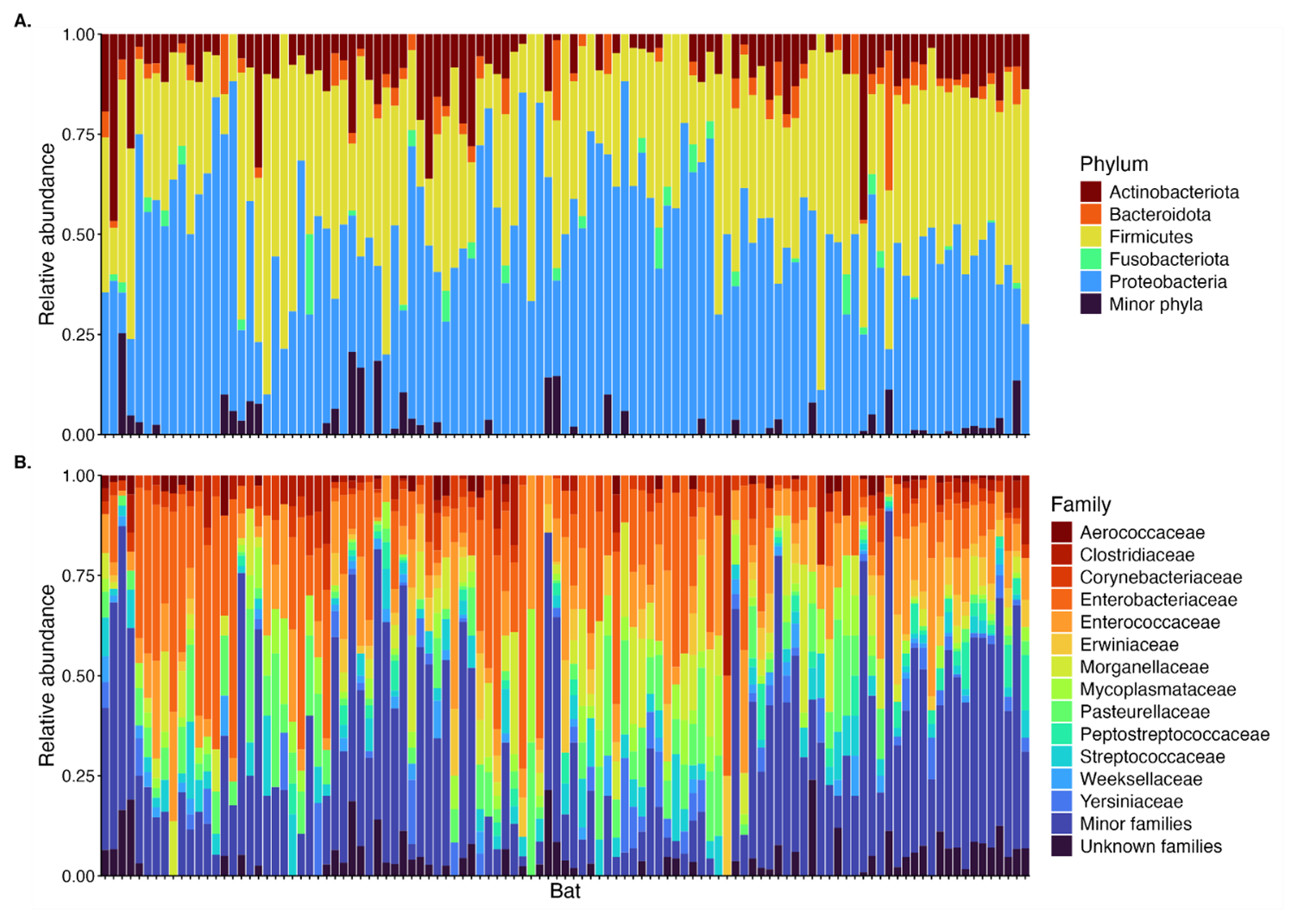
Figure 2Relative abundance of most common bacterial phyla (A) and families (B) found across all samples. Each bar represents an individual bat. Colors correspond to the bacterial phylum or family. “Minor phyla” (A) correspond to phyla found in <20 bats. “Minor families” (B) correspond to families found in <40 bats.
3.2 Factors influencing bacterial species richness and phylogenetic diversity
We found that there was no significant difference in either ASV richness based on sex (, p=0.28), age (t=0.62, p=0.55), or reproductive condition (chi-squared=5.9, p=0.12) (Fig. S4). We observed a similar pattern for phylogenetic diversity, again with no significant differences based on sex (, p=0.14), age (t=0.45, p=0.67), or reproductive condition (chi-squared=3.4, p=0.33) (Fig. S5). In addition, we found no correlation between either ASV richness (ρ=0.23, p=0.02) or phylogenetic diversity (ρ=0.09, p=0.33) and body condition (Figs. S6 and S7).
We found no association between land cover composition or configuration on ASV richness as the top model was the null model (Table S1 in the Supplement). Similarly, there was no clear association between phylogenetic diversity and land cover composition or configuration. While the model that best explained phylogenetic diversity included water cover at the landscape scale, this variable was not significant (β=1.1, 95 % CI: −0.58–2.7, p=0.22). All other models, including the null model, were within 2 AICc units of this model, and model weight was low (0.16) (Table S2).
3.3 Potential bacterial pathogens
We detected ASVs belonging to five potentially pathogenic bacterial genera: Bartonella, Campylobacter, Mycoplasma, Rickettsia, and Salmonella. Bartonella was represented by eight ASVs, one identified as Bartonella tamiae (although the 16S marker is generally not reliable for species-level identification for this genus) (Kosoy et al., 2012), and the others belonging to unknown species. Campylobacter was represented by a single ASV belonging to an unknown species. Mycoplasma was represented by seven ASVs, none identified to the species level. Rickettsia was represented by 10 ASVs, none identified to the species level. Salmonella was represented by 11 ASVs, three of which were identified as S. enterica.
Mycoplasma was detected at a strikingly high prevalence: 87 % (n=95) of bats at all but two roosts (n=15, 88 %), with 1–12 positive bats detected per roost. The other three genera were found at lower but still relatively high prevalence: 36 % (n=39) for Rickettsia, 34 % (n=37) for Salmonella, and 23 % (n=25) for Bartonella. Rickettsia and Salmonella were found in 59 % of surveyed roosts (n=10), with 1–10 bats per roost for Rickettsia and 1–8 for Salmonella, while bats with Bartonella were found in 53 % of roosts (n=9), with detection in 1–5 bats per roost. In contrast, Campylobacter was the most rarely detected pathogen, found in only four bats (4 %), all from different roosts. Co-occurrence of these potentially pathogenic genera in the same individuals was relatively common. We identified one bat in which all five genera co-occurred, seven bats with four genera, 23 with three genera, and 27 with a co-occurrence of two genera.
We did not find significant differences in prevalence for any of the five potential pathogens based on sex, age, or reproductive condition (see Table 1).
Table 1Summary of results of χ2 tests measuring differences in prevalence of ASVs belonging to the genera Bartonella, Campylobacter, Mycoplasma, Rickettsia, and Salmonella based on the sex, age, and reproductive condition of bats.

The model that best explained Bartonella occurrence was landscape-scale savanna splitting, but it was not significant (, 95 % CI: −12.0 to −0.43, p=0.17). Model weight was low compared to the other models (0.30), with several competing models (ΔAICc<2): landscape-scale sugarcane cover, landscape-scale savanna cover, and fine-scale savanna cover (Table S3).
The occurrence of Campylobacter, Mycoplasma, Rickettsia, and Salmonella were not well explained by any land cover composition or configuration variables; the model that best explained all of their occurrence was the null model (Tables S4–S7).
3.4 Bacterial community
The NMDS plots showed no clear clustering of the bacterial community based on sex, age, or reproductive status (Fig. S8).
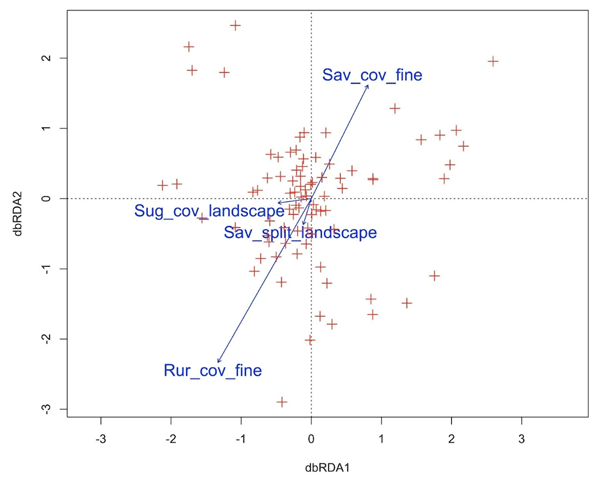
The global distance-based redundancy analysis (db-RDA) model based on Jaccard's distance was significant (p=0.001) but explained only a relatively small amount of the variation in community composition (15 %), while the spatial component explained 19 %, and 66 % of the variation remained unexplained. When controlling for the number of variables, the adjusted R2=0.03. The forward-selected db-RDA included four significant covariates: fine-scale rural cover (p=0.001), fine-scale savanna cover (p=0.001), landscape-scale sugarcane cover (p=0.003), and landscape-scale savanna splitting (p=0.03) (Fig. 3). However, the portion of variation explained was reduced to 6.7 %, but the adjusted R2 was only marginally affected (adjusted R2=0.026).

Figure 3Distance-based redundancy (db-RDA) plot based on Jaccard's distance showing the relationship between the land cover variables selected by forward selection: fine-scale rural cover (Rur_cov_fine), fine-scale savanna cover (Sav_cov_fine), landscape-scale sugarcane cover (Sug_cov_landscape), and landscape-scale savanna splitting (Sav_split_landscape), along with the bacterial communities within bats. Red crosses indicate bats. Together, all covariates described 6.7 % of the variation (adjusted R2=0.026).
Similarly, the db-RDA based on UniFrac distances was significant (p=0.001) but explained slightly less of the variation in phylogenetic community composition (14 %) compared to the ASV-based community composition. Meanwhile, the spatial component explained a slightly greater amount of the phylogenetic variation at 23 %, and 62 % remained unexplained. When controlling for the number of variables, the adjusted R2=0.03. The forward-selected model retained two variables, both of which were significant: fine-scale savanna cover (p=0.001) and landscape-scale water cover (p=0.04); however, the portion of variation explained was only 3.2 %, with an adjusted R2 of only =0.013.
4.1 Bacterial identification and occurrence
This is the first study of the microbiome of Mops pumilus. We found that this bat hosted a species-rich microbiome, with a total of 2249 ASVs identified in the full data set and up to 185 ASVs in a single individual bat. Our sample-based rarefaction curve indicated that this is a good representation of the diversity of the species' gut microbiome, at least within the geographical region, likely due to sampling depth and the relatively high number of individuals from a single species. The richness was comparable to what has been observed in other insectivorous species (Dietrich et al., 2017; Hughes et al., 2018), although other studies have found much greater species richness in Phyllostomid (Carrillo-Araujo et al., 2015) and Vespertilionid insectivores (Bennett et al., 2025). At the individual level, bacterial species richness was similar to what has been observed in other molossids in Africa, including M. bivitattus, belonging to the same genus (Lutz et al., 2019).
The most common phyla we identified were Proteobacteria, Firmicutes, and Actinobacteria, a pattern that differs from other mammals but appears to be general across bat species and geographic regions (Carrillo-Araujo et al., 2015; Dietrich et al., 2017; Hughes et al., 2018; Lutz et al., 2019; Nishida and Ochman, 2018). Similarly, the most commonly occurring bacterial families in M. pumilus – Enterobacteriaceae and Streptococcaceae – are also common in other bat species, including both insectivores and frugivores (Gerbáčová et al., 2020; Ingala et al., 2018; Li et al., 2018; Lutz et al., 2019; Popov et al., 2023).
We found high variability in the specific ASVs present within the microbiome of individuals, with most ASVs occurring in only a single bat. High levels of individual variability have previously been observed in bats (Alpízar et al., 2021; Banskar et al., 2016; Lutz et al., 2022; Núñez-Montero et al., 2021; Vengust et al., 2018), even for groups in captivity with the same diet and housing (Riopelle et al., 2024). Gut microbiomes based on fecal samples may be particularly susceptible to high levels of variation because they capture the effect of diet more than phylogeny or evolutionary history (Ingala et al., 2018). Mops pumilus is a generalist insectivore that feeds on a wide range of insect species (Bohmann et al., 2011; Bouchard, 1998). As diet may vary widely between individuals (Bohmann et al., 2011), it is not surprising that the diversity and identity of ASVs in the microbiome of M. pumilus would differ as well. While we hypothesize that the high levels of individual variation in gut microbiome reflect the high variability in diet observed in M. pumilus (Bohmann et al., 2011), we are unable to make a direct link between these factors. Further investigation directly correlating gut microbiome diversity and community composition with variation in diet could shed more light on these associations at the individual level within a species. In addition, while studies have shown that gut microbiome is dynamic and changes over time (Riopelle et al., 2024), we sampled each individual at a single point in time.
4.2 Factors influencing bacterial species richness, phylogenetic diversity, and community
We found no significant differences in either bacterial species richness, phylogenetic diversity, or community composition between males and females despite relatively balanced sample sizes (58 females vs. 45 males). Evidence from other studies is mixed, with some finding sex differences (Arellano-Hernández et al., 2024; Dietrich et al., 2018; Fleischer et al., 2024; Riopelle et al., 2024), while others did not (Lobato-Bailón et al., 2023; Núñez-Montero et al., 2021). We found no differences based on reproductive condition, although other studies have (Dietrich et al., 2018; Gaona et al., 2019; Phillips et al., 2012). This may be related to sample sizes, with only four pregnant and no lactating females captured. As we did not detect differences between the sexes, it is perhaps not surprising that bacterial richness, phylogenetic diversity, and composition did not vary between non-reproductive bats, scrotal males, and post-lactating females. Analyzing a larger sample size of pregnant and lactating female M. pumilus could yield different results. The lack of differences between adults and juveniles has also been found in other species, perhaps indicating a stable gut microbiome once young bats are weaned and able to forage themselves (Gaona et al., 2019; Hughes et al., 2018), although we again note a very small sample size for juveniles (n=9).
We found that land cover had no effect on species richness of the gut microbiome in M. pumilus. This is in contrast to other studies (Alpízar et al., 2021; Lobato-Bailón et al., 2023). Mops pumilus is highly tolerant of anthropogenic disturbance, for example, large numbers can be found roosting in buildings (Bouchard, 1998; Shapiro and Monadjem, 2016), and they forage extensively over sugarcane plantations (Noer et al., 2012; Shapiro et al., 2020). As their activity and foraging habits are not negatively impacted by anthropogenic land cover or degradation, their gut microbiomes may also be largely unaffected as well. The marginal effects of land cover that we did see on microbiome community composition were stronger at the fine scale than the landscape scale, despite the fact that M. pumilus has a large home range and can fly over 4 km from its roost nightly (Noer et al., 2012). However, the activity of this species is also best explained by both fine- and landscape-scale variables, depending on the season (Shapiro et al., 2020). The fact that we found effects at both scales on the gut microbiome may reflect the influences of both foraging activity at large scales and the finer-scale variables surrounding their roosts, where bats spend the majority of their time. However, as the effects were small, other variables associated with the local environment, including the local environmental microbiome (Bernardo-Cravo et al., 2020) or roost characteristics, may ultimately be more important than land cover composition (Lutz et al., 2022). Social interactions within each roost likely also have a strong effect on shaping the gut microbiome of the bats using it (Kolodny et al., 2019; Lebeuf-Taylor et al., 2025). However, due to the way we sampled (many roosts, relatively small sample sizes for each, uneven sample sizes between them), these effects are difficult to untangle within our data set. We would not expect kinship to be a factor within roosts to confound these results as M. pumilus disperse from their natal roosts at maturity, and therefore individuals at a single roost would not be more related to each other than those in different roosts (Bouchard, 1998; Mcwilliam, 1987). Complementary analyses, such as network approaches, could shed light on these patterns.
We note that our sampling strategy prioritized spatial coverage and variability in landscape metrics rather than temporal scale. We did not explicitly sample by season, instead sampling continuously from November to July, with a large proportion of bats captured in November and March to May, periods that are between the wet and dry season (Shapiro et al., 2020). Therefore, we cannot capture the effect of season on gut microbiome, which may influence microbiome due to reproductive cycles (Dietrich et al., 2016) or shifts in diet (Xiong et al., 2026). However, as M. pumilus do not migrate or hibernate (Bouchard, 1998; Monadjem et al., 2020), which drives seasonal changes in gut microbiome in other bats species (Shao et al., 2026; Víquez-R et al., 2021; Xiao et al., 2019), such shifts could likely be less pronounced in this species.
4.3 Potentially pathogenic bacteria
We detected ASVs belonging to five potentially pathogenic bacterial genera – Mycoplasma (88 %), Rickettsia (36 %), Salmonella (34 %), Bartonella (23 %), and Campylobacter (4 %) – with the first four in particular at high prevalence. All five of these genera are relatively common in bats worldwide (Ferreira et al., 2024; Mühldorfer, 2013; Szentivanyi et al., 2023). Owing to the use of fecal samples, we could not determine whether any of the five potentially pathogenic genera that we detected were in fact being shed by infected bats. Screening of other sample types, particularly blood or saliva, could provide evidence of any potential infection and shedding (Dietrich et al., 2016, 2018). Nevertheless, as we did not detect any signs of disease, ill health, or poor body condition, it is more likely that bats were asymptomatic carriers or that these bacteria originated in their insect prey, rather than being carried by the bats themselves. While bacterial disease caused by Borrelia (Evans et al., 2009), Staphylococcus aureus (Weinberg et al., 2022, 2026), and Yersinia enterocolita (Imnadze et al., 2020) have been reported in bats, we are not aware of studies showing a direct pathogenic effect of Mycoplasma, Rickettsia, Salmonella, Bartonella, or Campylobacter specifically in bats. However, previous studies have found correlations between the presence of potentially pathogenic bacterial genera and enteric viruses, with authors hypothesizing that the viral infections altered gut microbiome, potentially leading to a state of dysbiosis, and left hosts with worse body condition and more vulnerable to colonization by these bacteria (Melville et al., 2025; Wasimuddin et al., 2018).
Mycoplasma in particular was detected at a strikingly high prevalence of 88 %, and the most common ASV across individual bats belonged to this genus as well. While this is higher than the prevalence reported in most studies (Becker et al., 2024; Di Cataldo et al., 2020; Mascarelli et al., 2014; Volokhov et al., 2023; Wang et al., 2023), it is comparable to results from Spain where Mycoplasma was detected in 97 % (n=31) of sampled Miniopterus schreibersii and Myotis capaccinii (Millán et al., 2015). This high prevalence suggests that the Mycoplasma we detected are unlikely to be pathogenic. The seven distinct Mycoplasma ASVs that we identified demonstrate a high diversity of this genus within the M. pumilus population, which is in line with other studies (Millán et al., 2015; Volokhov et al., 2023; Wang et al., 2023). While we found no indication of illness in the bats, this genus can be asymptomatic in certain individuals in species in which it is pathogenic (Dawood et al., 2022). Nevertheless, Mycoplasma are considered emerging pathogens that have caused mortality in other wildlife species (Hartup et al., 2001; Tardy et al., 2012). Further, Pteropus species in New Caledonia have been shown to be reservoirs of M. haemohominis, and cases in humans have been epidemiologically linked to contact with these bats (Descloux et al., 2021). Surveillance and vigilance regarding this genus in M. pumilus may be warranted.
Bartonella and Rickettsia are both vector borne (Kosoy et al., 2012; Weinert et al., 2009) and common across bat species worldwide (Stuckey et al., 2017; Szentivanyi et al., 2023). Both genera have previously been detected in insectivorous miniopterid, rhinolophid, and vespertilionid bats, along with the frugivorous pteropodid Rousettus aegyptiacus in South Africa as well as Nycteris thebaica in Eswatini (Dietrich et al., 2016, 2017). While the four samples from M. pumilus in Dietrich et al. (2016) were all negative, this is not unexpected considering the small sample size. Bartonella is often associated with the bat fly ectoparasites in the families Streblidae and Nycteribiidae (do Amaral et al., 2018; Billeter et al., 2012; Brook et al., 2015; Dietrich et al., 2016; Kamani et al., 2014; Leulmi et al., 2016; Veikkolainen et al., 2014), and to a lesser extent fleas, ticks, and mites (Hornok et al., 2019; Leulmi et al., 2016; Reeves et al., 2007; Veikkolainen et al., 2014). Rickettsia is typically found in ticks collected from bats (Cicuttin et al., 2017; Hornok et al., 2019; Tompa et al., 2023; Zhao et al., 2020), although it has been detected in bat flies as well (do Amaral et al., 2018). However, this genus is also a common insect endosymbiont (Weinert et al., 2009). The detection of Rickettsia in particular may be due to feeding on infected insects or insects that had fed on infected plants, or the consumption of ticks they groomed off. These scenarios could all lead to the detection of Rickettsia in feces, without causing infection in the bats themselves.
Salmonella and Campylobacter are both enteric pathogens that may cause diarrheal disease in humans and other animals (Coburn et al., 2007; Man, 2011). When detected in bats, Salmonella is usually found at low prevalence ranging from <1 % to 12 % (Adesiyun et al., 2009; Bernardo Reyes et al., 2011; Ferreira et al., 2021; Islam et al., 2013; Mühldorfer, 2013), although a study in Nigeria found much higher prevalence at 72 % (Aladejana et al., 2025). It has been suggested that these bacteria are transmitted to bats via contaminated water (Dutta et al., 2025; Islam et al., 2013). It is unclear whether this might be plausible in northeastern Eswatini, although a study in a neighboring province of South Africa did detect Salmonella in groundwater (Mpenyana-Monyatsi et al., 2012). While Campylobacter is generally associated with wild birds and poultry, recent studies have detected novel, emerging species in wild mammals (Beisele et al., 2011; Goldman et al., 2011; Man, 2011). In bats, this genus has been rarely detected (Mühldorfer, 2013). In the Philippines, Campylobacter was found in 7.5 % of sampled Rousettus amplexicaudatus and was one of the most relatively abundant genera (Hatta et al., 2016). It was also detected at 3 % in six Vespertilionid bat species in the Netherlands (Hazeleger et al., 2018). In Finland, Campylobacter prevalence was higher in terrestrial small mammals found near livestock farms compared to natural habitats (Olkkola et al., 2021). In contrast, we did not find any association with Campylobacter prevalence and land cover, likely due to the fact that we detected this genus in only four bats from four very different sites.
In conclusion, we find that M. pumilus harbors a diverse gut microbiome marked by high variability between individuals. The limited effects of land cover composition and configuration metrics may be indicative of this species' generally adaptability to anthropogenic disturbance. The detection of potentially pathogenic bacteria is cause for vigilance for the health of both M. pumilus and other species, including humans, although there is currently no evidence that they cause illness in their chiropteran hosts, or can infect humans or other animals. Nevertheless, continued research and surveillance of the M. pumilus microbiome can contribute to both the health and conservation of this species and those they might interact with from a One Health perspective.
All code and scripts are available at https://github.com/jtshapiro/Mops_pumilus_Gut_microbiomes and https://doi.org/10.5281/zenodo.18614739 (Shapiro, 2026).
Raw sequencing data are deposited in the Sequence Read Archive, the National Center for Biotechnology Information (BioProject: PRJNA1420193). All other data files are available at https://github.com/jtshapiro/Mops_pumilus_Gut_microbiomes and https://doi.org/10.5281/zenodo.18614739 (Shapiro, 2026).
The supplement related to this article is available online at https://doi.org/10.5194/we-26-223-2026-supplement.
JTS, RAM, AJH, and AM conceived the study. JTS and AM collected samples. JTS, IBN, and LR conducted the laboratory work. JTS and CB carried out bioinformatic analyses. JTS carried out statistical analyses. JTS prepared the paper, with contributions from all authors.
The contact author has declared that none of the authors has any competing interests.
Publisher's note: Copernicus Publications remains neutral with regard to jurisdictional claims made in the text, published maps, institutional affiliations, or any other geographical representation in this paper. The authors bear the ultimate responsibility for providing appropriate place names. Views expressed in the text are those of the authors and do not necessarily reflect the views of the publisher.
We would like to thank Hervé Echecolonea, Mnqobi Mamba, Muzi Sibiya, Bonginkhosi Gumbi, and Alexandra Howard for assistance in the field. We are grateful to Mandla Motsa, Smart Shabangu, Tal Fineberg, and the staff at the Mbuluzi Game Reserve; Thea Litschka-Koen, Clifton Koen; Alan Howland and staff at the IYSIS Cattle and Game Ranch; and the Savanna Research Center, Kim Roques, and All Out Africa for help with logistics. We thank the residents in northeastern Eswatini who allowed us to capture bats in their villages and homes.
This material is based upon work supported by the National Science Foundation Graduate Research Fellowship under Grant No. DGE-1315138 (JTS), a National Science Foundation Graduate Research Opportunities Worldwide grant (JTS), a Student Research Grant from Bat Conservation International (JTS), a National Geographic Young Explorer's Grant 9635-14 (JTS), a grant from The Explorers Club Exploration Fund – Mamont Scholars Program (JTS), a grant from the American-Scandinavian Foundation – Amanda E. Roleson Fund (JTS), and a grant from the University of Florida Biodiversity Institute Fellowship (JTS). (submitted version).
This paper was edited by Erinne Stirling and reviewed by Eleonore Lebeuf-Taylor and two anonymous referees.
Adesiyun, A. A., Stewart-Johnson, A., and Thompson, N. N.: Isolation of enteric pathogens from bats in Trinidad, J. Wildlife Dis., 45, 952–961, https://doi.org/10.7589/0090-3558-45.4.952, 2009.
Aladejana, O. M., Oluduro, A. O., Ogunlade, A. O., Babatunde, S. K., and Sanni, O. T.: Isolation, molecular identification and antibiotics susceptibility of Salmonella serotypes isolates from bats in Osun State, Nigeria, Vegetos, 38, 668–675, https://doi.org/10.1007/s42535-023-00791-2, 2025.
Aleman, J. C., Blarquez, O., and Staver, C. A.: Land-use change outweighs projected effects of changing rainfall on tree cover in sub-Saharan Africa, Glob. Change Biol., 22, 3013–3025, https://doi.org/10.1111/gcb.13299, 2016.
Alpízar, P., Risely, A., Tschapka, M., and Sommer, S.: Agricultural fast food: Bats feeding in banana monocultures are heavier but have less diverse gut microbiota, Front. Ecol. Evol., 9, https://doi.org/10.3389/fevo.2021.746783, 2021.
Amato, K. R., Yeoman, C. J., Kent, A., Righini, N., Carbonero, F., Estrada, A., Gaskins, H. R., Stumpf, R. M., Yildirim, S., Torralba, M., Gillis, M., Wilson, B. A., Nelson, K. E., White, B. A., and Leigh, S. R.: Habitat degradation impacts black howler monkey (Alouatta pigra) gastrointestinal microbiomes, ISME J., 7, 1344–1353, https://doi.org/10.1038/ismej.2013.16, 2013.
Arellano-Hernández, H. D., Montes-Carreto, L. M., Guerrero, J. A., and Martinez-Romero, E.: The fecal microbiota of the mouse-eared bat (Myotis velifer) with new records of microbial taxa for bats, PLoS One, 19, e0314847, https://doi.org/10.1371/journal.pone.0314847, 2024.
Bäckhed, F., Ding, H., Wang, T., Hooper, L. V., Koh, G. Y., Nagy, A., Semenkovich, C. F., and Gordon, J. I.: The gut microbiota as an environmental factor that regulates fat storage, P. Natl. Acad. Sci. USA, 101, 15718–15723, https://doi.org/10.1073/pnas.0407076101, 2004.
Bailey, K. M., McCleery, R. A., Binford, M. W., and Zweig, C.: Land-cover change within and around protected areas in a biodiversity hotspot, J. Land Use Sci., 1–23, https://doi.org/10.1080/1747423X.2015.1086905, 2015.
Banskar, S., Mourya, D. T., and Shouche, Y. S.: Bacterial diversity indicates dietary overlap among bats of different feeding habits, Microbiol. Res., 182, 99–108, https://doi.org/10.1016/j.micres.2015.10.006, 2016.
Barelli, C., Albanese, D., Donati, C., Pindo, M., Dallago, C., Rovero, F., Cavalieri, D., Tuohy, K. M., Hauffe, H. C., and De Filippo, C.: Habitat fragmentation is associated to gut microbiota diversity of an endangered primate: implications for conservation, Sci. Rep., 5, 14862, https://doi.org/10.1038/srep14862, 2015.
Barnes, C. J., Rasmussen, L., Asplund, M., Knudsen, S. W., Clausen, M.-L., Agner, T., and Hansen, A. J.: Comparing DADA2 and OTU clustering approaches in studying the bacterial communities of atopic dermatitis, J. Med. Microbiol., 69, 1293–1302, https://doi.org/10.1099/jmm.0.001256, 2020.
Barton, K.: MuMIn: Multi-model inference. R package version 1.40.0, https://CRAN.R-project.org/package=MuMIn (last access: 28 December 2025), 2017.
Bates, D., Mächler, M., Bolker, B., and Walker, S.: Fitting linear mixed-effects models using lme4, J. Stat. Softw., 67, 1–48, https://doi.org/10.18637/jss.v067.i01, 2015.
Becker, D. J., Dyer, K. E., Lock, L. R., Olbrys, B. L., Pladas, S. A., Sukhadia, A. A., Demory, B., Nunes Batista, J. M., Pineda, M., Simmons, N. B., Adams, A. M., Frick, W. F., O'Mara, M. T., and Volokhov, D. V.: Geographically widespread and novel hemotropic mycoplasmas and bartonellae in Mexican free-tailed bats and sympatric North American bat species, mSphere, 10, e00116-24, https://doi.org/10.1128/msphere.00116-24, 2024.
Beisele, M., Shen, Z., Parry, N., Mobley, M., Taylor, N. S., Buckley, E., Abedin, M. Z., Dewhirst, F. E., and Fox, J. G.: Helicobactermarmotae and novel Helicobacter and Campylobacter species isolated from the livers and intestines of prairie dogs, J. Med. Microbiol., 60, 1366–1374, https://doi.org/10.1099/jmm.0.032144-0, 2011.
Bennett, A. J., Suski, C. D., and O'Keefe, J. M.: Molecular epidemiology of Eimeria spp. parasites and the faecal microbiome of Indiana bats (Myotis sodalis): A non-invasive, multiplex metabarcode survey of an endangered species, Microb. Genom., 11, 001358, https://doi.org/10.1099/mgen.0.001358, 2025.
Bernardo-Cravo, A. P., Schmeller, D. S., Chatzinotas, A., Vredenburg, V. T., and Loyau, A.: Environmental factors and host microbiomes shape host–pathogen dynamics, Trends Parasitol., 36, 616–633, https://doi.org/10.1016/j.pt.2020.04.010, 2020.
Bernardo Reyes, A. W., Rovira, H. G., Masangkay, J. S., Ramirez, T. J., Yoshikawa, Y., and Baticados, W. N.: Polymerase Chain Reaction assay and conventional isolation of Salmonella spp. from Philippine bats, Acta Sci. Vet., 39, 1, https://www.redalyc.org/pdf/2890/289022244008.pdf (last access: 18 September 2026), 2011.
Biagi, E., Franceschi, C., Rampelli, S., Severgnini, M., Ostan, R., Turroni, S., Consolandi, C., Quercia, S., Scurti, M., Monti, D., Capri, M., Brigidi, P., and Candela, M.: Gut microbiota and extreme longevity, Curr. Biol., 26, 1480–1485, https://doi.org/10.1016/j.cub.2016.04.016, 2016.
Billeter, S. A., Hayman, D. T. S., Peel, A. J., Baker, K., Wood, J. L. N., Cunningham, A., Suu-Ire, R., Dittmar, K., and Kosoy, M. Y.: Bartonella species in bat flies (Diptera: Nycteribiidae) from western Africa, Parasitology, 139, 324–329, https://doi.org/10.1017/S0031182011002113, 2012.
Blanchet, F. G., Legendre, P., and Borcard, D.: Forward selection of explanatory variables, Ecology, 89, 2623–2632, https://doi.org/10.1890/07-0986.1, 2008.
Bodenhofer, U., Bonatesta, E., Horejš-Kainrath, C., and Hochreiter, S.: msa: an R package for multiple sequence alignment, Bioinformatics, 31, 3997–3999, https://doi.org/10.1093/bioinformatics/btv494, 2015.
Bohmann, K., Monadjem, A., Noer, C. L., Rasmussen, M., Zeale, M. R. K., Clare, E., Jones, G., Willerslev, E., and Gilbert, M. T. P.: Molecular diet analysis of two African free-tailed bats (Molossidae) using high throughput sequencing, PLoS One, 6, e21441, https://doi.org/10.1371/journal.pone.0021441, 2011.
Borcard, D., Legendre, P., and Drapeau, P.: Partialling out the spatial component of ecological variation, Ecology, 73, 1045–1055, https://doi.org/10.2307/1940179, 1992.
Bouchard, S.: Chaerephon pumilus, Mamm. Species, 574, 1–6, 1998.
Brook, C. E., Bai, Y., Dobson, A. P., Osikowicz, L. M., Ranaivoson, H. C., Zhu, Q., Kosoy, M. Y., and Dittmar, K.: Bartonella spp. in fruit bats and blood-feeding ectoparasites in Madagascar, PLoS Neglect. Trop. D., 9, e0003532, https://doi.org/10.1371/journal.pntd.0003532, 2015.
Burgin, C. J., Zijlstra, J. S., Becker, M. A., Handika, H., Alston, J. M., Widness, J., Liphardt, S., Huckaby, D. G., and Upham, N. S.: How many mammal species are there now? Updates and trends in taxonomic, nomenclatural, and geographic knowledge, J. Mammal., 106, 1082–1117, https://doi.org/10.1093/jmammal/gyaf047, 2025.
Burnham, K. P. and Anderson, D. R.: Model Selection and Multimodel Inference: A Practical Information-Theoretic Approach, Springer-Verlag, New York, 488 pp., https://doi.org/10.1007/b97636, 2002.
Bustin, S. A., Benes, V., Garson, J. A., Hellemans, J., Huggett, J., Kubista, M., Mueller, R., Nolan, T., Pfaffl, M. W., Shipley, G. L., Vandesompele, J., and Wittwer, C. T.: The MIQE guidelines: Minimum information for publication of quantitative real-time PCR experiments, Clin. Chem., 55, 611–622, https://doi.org/10.1373/clinchem.2008.112797, 2009.
Callahan, B. J., McMurdie, P. J., Rosen, M. J., Han, A. W., Johnson, A. J. A., and Holmes, S. P.: DADA2: High-resolution sample inference from Illumina amplicon data, Nat. Methods, 13, 581–583, https://doi.org/10.1038/nmeth.3869, 2016.
Carmody, R. N., Gerber, G. K., Luevano, J. M., Gatti, D. M., Somes, L., Svenson, K. L., and Turnbaugh, P. J.: Diet dominates host genotype in shaping the murine gut microbiota, Cell Host Microbe, 17, 72–84, https://doi.org/10.1016/J.CHOM.2014.11.010, 2015.
Carrillo-Araujo, M., Taş, N., Alcántara-Hernández, R. J., Gaona, O., Schondube, J. E., Medellín, R. A., Jansson, J. K., and Falcón, L. I.: Phyllostomid bat microbiome composition is associated to host phylogeny and feeding strategies, Front. Microbiol., 6, 447, https://doi.org/10.3389/fmicb.2015.00447, 2015.
Chambers, C. L., Cushman, S. A., Medina-Fitoria, A., Martínez-Fonseca, J., and Chávez-Velásquez, M.: Influences of scale on bat habitat relationships in a forested landscape in Nicaragua, Landscape Ecol., 31, 1299–1318, https://doi.org/10.1007/s10980-016-0343-4, 2016.
Chang, C.-W., Huang, B.-H., Lin, S.-M., Huang, C.-L., and Liao, P.-C.: Changes of diet and dominant intestinal microbes in farmland frogs, BMC Microbiol., 16, 33, https://doi.org/10.1186/s12866-016-0660-4, 2016.
Chao, A.: Estimating the population size for capture-recapture data with unequal catchability, Biometrics, 43, 783–791, 1987.
Charif, D. and Lobry, J.: SeqinR 1.0-2: a contributed package to the R project for statistical computing devoted to biological sequences retrieval and analysis, in: Structural approaches to sequence evolution: Molecules, networks, populations, series Biological and Medical Physics, Biomedical Engineering, edited by: Bastolla, U., Porto, M., Roman, H., and Vendruscolo, M., Springer Verlag, New York, 207–232, ISBN 978-3-540-35305-8, 2007.
Cicuttin, G. L., De Salvo, M. N., La Rosa, I., and Dohmen, F. E. G.: Neorickettsia risticii, Rickettsia sp. and Bartonella sp. in Tadarida brasiliensis bats from Buenos Aires, Argentina, Comp. Immunol. Microb., 52, 1–5, https://doi.org/10.1016/j.cimid.2017.04.004, 2017.
Claesson, M. J., Jeffery, I. B., Conde, S., Power, S. E., O'Connor, E. M., Cusack, S., Harris, H. M. B., Coakley, M., Lakshminarayanan, B., O'Sullivan, O., Fitzgerald, G. F., Deane, J., O'Connor, M., Harnedy, N., O'Connor, K., O'Mahony, D., Van Sinderen, D., Wallace, M., Brennan, L., Stanton, C., Marchesi, J. R., Fitzgerald, A. P., Shanahan, F., Hill, C., Ross, R. P., and O'Toole, P. W.: Gut microbiota composition correlates with diet and health in the elderly, Nature, 488, 178–185, https://doi.org/10.1038/nature11319, 2012.
Coburn, B., Grassl, G. A., and Finlay, B. B.: Salmonella, the host and disease: A brief review, Immunol. Cell Biol., 85, 112–118, https://doi.org/10.1038/sj.icb.7100007, 2007.
Daszak, P., Cunningham, A. A. A., and Hyatt, A. D. D.: Anthropogenic environmental change and the emergence of infectious diseases in wildlife, Acta Trop., 78, 103–116, https://doi.org/10.1016/s0001-706x(00)00179-0, 2001.
Dawood, A., Algharib, S. A., Zhao, G., Zhu, T., Qi, M., Delai, K., Hao, Z., Marawan, M. A., Shirani, I., and Guo, A.: Mycoplasmas as host pantropic and specific pathogens: Clinical implications, gene transfer, virulence factors, and future perspectives, Front. Cell. Infect. Mi., 12, 855731, https://doi.org/10.3389/fcimb.2022.855731, 2022.
Descloux, E., Mediannikov, O., Gourinat, A.-C., Colot, J., Chauvet, M., Mermoud, I., Desoutter, D., Cazorla, C., Klement-Frutos, E., Antonini, L., Levasseur, A., Bossi, V., Davoust, B., Merlet, A., Goujart, M.-A., Oedin, M., Brescia, F., Laumond, S., Fournier, P.-E., and Raoult, D.: Flying fox hemolytic fever, description of a new zoonosis caused by Candidatus Mycoplasma haemohominis, Clin. Infect. Dis., 73, e1445–e1453, https://doi.org/10.1093/cid/ciaa1648, 2021.
Di Cataldo, S., Kamani, J., Cevidanes, A., Msheliza, E. G., and Millán, J.: Hemotropic mycoplasmas in bats captured near human settlements in Nigeria, Comp. Immunol. Microb., 70, 101448, https://doi.org/10.1016/j.cimid.2020.101448, 2020.
Dietrich, M., Tjale, M. A., Weyer, J., Kearney, T., Seamark, E. C. J., Nel, L. H., Monadjem, A., and Markotter, W.: Diversity of Bartonella and Rickettsia spp. in bats and their blood-feeding ectoparasites from South Africa and Swaziland, PLoS One, 11, e0152077, https://doi.org/10.1371/journal.pone.0152077, 2016.
Dietrich, M., Kearney, T., Seamark, E. C. J., and Markotter, W.: The excreted microbiota of bats: Evidence of niche specialisation based on multiple body habitats, FEMS Microbiol. Lett., 364, fnw284, https://doi.org/10.1093/femsle/fnw284, 2017.
Dietrich, M., Kearney, T., Seamark, E. C. J., Paweska, J. T., and Markotter, W.: Synchronized shift of oral, faecal and urinary microbiotas in bats and natural infection dynamics during seasonal reproduction, Roy. Soc. Open Sci., 5, 180041, https://doi.org/10.1098/rsos.180041, 2018.
do Amaral, R. B., Lourenço, E. C., Famadas, K. M., Garcia, A. B., Machado, R. Z., and André, M. R.: Molecular detection of Bartonella spp. and Rickettsia spp. in bat ectoparasites in Brazil, PLoS One, 13, e0198629, https://doi.org/10.1371/journal.pone.0198629, 2018.
Dutta, P., Uddin, Md. H., Islam, S., Islam, M., Hossain, M. S., Sagor, M. S., Rumi, M. A., Rahman, M. K., FLora, M. S., Samad, M. A., Forwood, J. K., Epstein, J. H., Chowdhury, S., and Islam, A.: Epidemiology and ecology of multidrug resistance (MDR) Salmonella in fruit bats in Bangladesh, The 20th International Bat Research Conference (IBRC) 2025, 171–171, https://researchoutput.csu.edu.au/en/publications/epidemiology-and-ecology-of-multidrug-resistance-mdr-salmonella-i/ (last access: 18 September 2026), 2025.
Edgar, R. C.: UNBIAS: An attempt to correct abundance bias in 16S sequencing, with limited success, bioRxiv [preprint], https://doi.org/10.1101/124149, 2017.
Elbrecht, V. and Leese, F.: Can DNA-based ecosystem assessments quantify species abundance? Testing primer bias and biomass–sequence relationships with an innovative metabarcoding protocol, PLoS One 10, e0130324, https://doi.org/10.1371/journal.pone.0130324, 2015.
Ethier, K. and Fahrig, L.: Positive effects of forest fragmentation, independent of forest amount, on bat abundance in eastern Ontario, Canada, Landscape Ecol., 26, 865–876, https://doi.org/10.1007/s10980-011-9614-2, 2011.
Evans, N. J., Bown, K. J., Timofte, D., Simpson, V. R., and Birtles, R. J.: Fatal borreliosis in bat caused by relapsing fever spirochete, United Kingdom, Emerg. Infect. Dis., 15, 1331–1333, https://doi.org/10.3201/eid1508.090475, 2009.
Fackelmann, G., Gillingham, M. A. F., Schmid, J., Heni, A. C., Wilhem, K., Schwensow, N., and Sommer, S.: Human encroachment into wildlife gut microbiomes, Commun. Biol., 4, 800, https://doi.org/10.1038/s42003-021-02315-7, 2021.
Fahrig, L.: Effects of habitat fragmentation on biodiversity, Annu. Rev. Ecol. Evol. S., 34, 487–515, https://doi.org/10.1146/annurev.ecolsys.34.011802.132419, 2003.
Faith, D. P.: Conservation evaluation and phylogenetic diversity, Biol. Conserv., 61, 1–10, https://doi.org/10.1016/0006-3207(92)91201-3, 1992.
Feddema, J. J., Oleson, K. W., Bonan, G. B., Mearns, L. O., Buja, L. E., Meehl, G. A., and Washington, W. M.: The importance of land-cover change in simulating future climates, Science, 310, 1674–1678, https://doi.org/10.1126/science.1118160, 2005.
Ferreira, A. C. R., Vieira, T. M., da Costa Custódio, D. A., Melo, M. N., Gontijo, C. M. F., Lage, A. P., and Dorneles, E. M. S.: Cross-sectional study on Brucella spp., Leptospira spp. and Salmonella spp. in bats from Montes Claros, Minas Gerais, Brazil, Comp. Immunol. Microb., 78, 101692, https://doi.org/10.1016/j.cimid.2021.101692, 2021.
Ferreira, A. C. R., Colocho, R. A. B., Pereira, C. R., Veira, T. M., Gregorin, R., Lage, A. P., and Dorneles, E. M. S.: Zoonotic bacterial pathogens in bats samples around the world: A scoping review, Prev. Vet. Med., 225, 106135, https://doi.org/10.1016/j.prevetmed.2024.106135, 2024.
Ficetola, G. F., Pansu, J., Elie Bonin, A., Coissac, E., Giguet-Covex, C., De Barba, M., Gielly, L., Lopes, C. M., and Boyer, E. E.: Replication levels, false presences and the estimation of the presence/absence from eDNA metabarcoding data, Mol. Ecol. Resour., 15, 543–556, https://doi.org/10.1111/1755-0998.12338, 2015.
Fischer, J. and Lindenmayer, D. B.: Beyond fragmentation: The continuum model for fauna research and conservation in human-modified landscapes, Oikos, 112, 473–480, https://doi.org/10.1111/j.0030-1299.2006.14148.x, 2006.
Fleischer, R., Jones, C., Ledezma-Campos, P., Czirják, G. Á., Sommer, S., Gillespie, T. R., and Vicente-Santos, A.: Gut microbial shifts in vampire bats linked to immunity due to changed diet in human disturbed landscapes, Sci. Total Environ., 907, 167815, https://doi.org/10.1016/j.scitotenv.2023.167815, 2024.
Frick, W. F., Kingston, T., and Flanders, J.: A review of the major threats and challenges to global bat conservation, Ann. NY Acad. Sci., 1469, 5–25, https://doi.org/10.1111/nyas.14045, 2020.
Frøslev, T. G., Kjøller, R., Bruun, H. H., Ejrnæs, R., Brunbjerg, A. K., Pietroni, C., and Hansen, A. J.: Algorithm for post-clustering curation of DNA amplicon data yields reliable biodiversity estimates, Nat. Commun., 8, 1188, https://doi.org/10.1038/s41467-017-01312-x, 2017.
Gaona, O., Gómez-Acata, E. S., Cerqueda-García, D., Neri-Barrios, C. X., and Falcón, L. I.: Fecal microbiota of different reproductive stages of the central population of the lesser-long nosed bat, Leptonycteris yerbabuenae, PLoS One, 14, e0219982, https://doi.org/10.1371/journal.pone.0219982, 2019.
Ge, X., Li, Y., Yang, X., Zhang, H., Zhou, P., Zhang, Y., and Shi, Z.: Metagenomic analysis of viruses from bat fecal samples reveals many novel viruses in insectivorous bats in China, J. Virol., 86, 4620–4630, https://doi.org/10.1128/jvi.06671-11, 2012.
Geldenhuys, M., Mortlock, M., Weyer, J., Bezuidt, O., Seamark, E. C. J., Kearney, T., Gleasner, C., Erkkila, T. H., Cui, H., and Markotter, W.: A metagenomic viral discovery approach identifies potential zoonotic and novel mammalian viruses in Neoromicia bats within South Africa, PLoS One, 13, e0194527, https://doi.org/10.1371/journal.pone.0194527, 2018.
Gerbáčová, K., Maliničová, L., Kisková, J., Maslišová, V., Uhrin, M., and Pristaš, P.: The faecal microbiome of building-dwelling insectivorous bats (Myotis myotis and Rhinolophus hipposideros) also Contains antibiotic-resistant bacterial representatives, Curr. Microbiol., 77, 2333–2344, https://doi.org/10.1007/s00284-020-02095-z, 2020.
Goldman, C. G., Matteo, M. J., Loureiro, J. D., Almuzara, M., Barberis, C., Vay, C., Catalano, M., Heredia, S. R., Mantero, P., Boccio, J. R., Zubillaga, M. B., Cremaschi, G. A., Solnick, J. V., Perez-Perez, G. I., and Blaser, M. J.: Novel gastric helicobacters and oral campylobacters are present in captive and wild cetaceans, Vet. Microbiol., 152, 138–145, https://doi.org/10.1016/j.vetmic.2011.04.023, 2011.
Goodrich, J. K., Waters, J. L., Poole, A. C., Sutter, J. L., Koren, O., Blekhman, R., Beaumont, M., Van Treuren, W., Knight, R., Bell, J. T., Spector, T. D., Clark, A. G., and Ley, R. E.: Human genetics shape the gut microbiome, Cell, 159, 789–799, https://doi.org/10.1016/J.CELL.2014.09.053, 2014.
Handy, R. D., Clark, N. J., Hutt, L. P., and Bescós, R.: The microbiomes of wildlife and chemical pollution: Status, knowledge gaps and challenges, Current Opinion in Toxicology, 36, 100428, https://doi.org/10.1016/j.cotox.2023.100428, 2023.
Hansen, C. H. F., Krych, L., Nielsen, D. S., Vogensen, F. K., Hansen, L. H., Sørensen, S. J., Buschard, K., and Hansen, A. K.: Early life treatment with vancomycin propagates Akkermansia muciniphila and reduces diabetes incidence in the NOD mouse, Diabetologia, 55, 2285–2294, https://doi.org/10.1007/s00125-012-2564-7, 2012.
Harrell Jr., F. E.: Hmisc: Harrell Miscellaneous. R package version 3.0-12, https://cran.r-project.org/web/packages/Hmisc/index.html (last access: 28 December 2025), 2006.
Hartup, B. K., Bickal, J. M., Dhondt, A. A., Ley, D. H., and Kollias, G. V.: Dynamics of conjunctivitis and Mycoplasma gallisepticum infections in house Finches, Auk, 118, 327–333, https://doi.org/10.1093/auk/118.2.327, 2001.
Hatta, Y., Omatsu, T., Tsuchiaka, S., Katayama, Y., Taniguchi, S., Masangkay, J. S., Puentespina Jr., R., Eres, E., Cosico, E., Une, Y., Yoshikawa, Y., Maeda, K., Kyuwa, S., and Mizutani, T.: Detection of Campylobacter jejuni in rectal swab samples from Rousettus amplexicaudatus in the Philippines, J. Vet. Med. Sci., 78, 1347–1350, https://doi.org/10.1292/jvms.15-0621, 2016.
Hayman, D. T. S.: Bats as viral reservoirs, Ann. Rev. Virol., 3, 77–99, https://doi.org/10.1146/annurev-virology-110615-042203, 2016.
Hazeleger, W. C., Jacobs-Reitsma, W. F., Lina, P. H. C., de Boer, A. G., Bosch, T., van Hoek, A. H. A. M., and Beumer, R. R.: Wild, insectivorous bats might be carriers of Campylobacter spp., PLoS One, 13, e0190647, https://doi.org/10.1371/journal.pone.0190647, 2018.
Hijmans, R. J.: geosphere: Spherical trigonometry, https://cran.r-project.org/web/packages/geosphere/index.html (last access: 18 September 2026), 2017.
Hornok, S., Szőke, K., Meli, M. L., Sándor, A. D., Görföl, T., Estók, P., Wang, Y., Tu, V. T., Kováts, D., Boldogh, S. A., Corduneanu, A., Sulyok, K. M., Gyuranecz, M., Kontschán, J., Takács, N., Halajian, A., Epis, S., and Hofmann-Lehmann, R.: Molecular detection of vector-borne bacteria in bat ticks (Acari: Ixodidae, Argasidae) from eight countries of the Old and New Worlds, Parasit. Vector., 12, 50, https://doi.org/10.1186/s13071-019-3303-4, 2019.
Hughes, G. M., Leech, J., Puechmaille, S. J., Lopez, J. V., and Teeling, E. C.: Is there a link between aging and microbiome diversity in exceptional mammalian longevity?, PeerJ, 6, e4174, https://doi.org/10.7717/peerj.4174, 2018.
Imnadze, T., Natradze, I., Zhgenti, E., Malania, L., Abazashvili, N., Sidamonidze, K., Khmaladze, E., Zakalashvili, M., Imnadze, P., Arner, R. J., Motin, V., and Kosoy, M.: Identification of a novel Yersinia enterocolitica strain from bats in association with a bat die-off that occurred in Georgia (Caucasus), Microorganisms, 8, 1000, https://doi.org/10.3390/microorganisms8071000, 2020.
Ingala, M. R., Simmons, N. B., Wultsch, C., Krampis, K., Speer, K. A., and Perkins, S. L.: Comparing microbiome sampling methods in a wild mammal: Fecal and intestinal samples record different signals of host ecology, evolution, Front. Microbiol., 9, 803, https://doi.org/10.3389/fmicb.2018.00803, 2018.
Ingala, M. R., Becker, D. J., Bak Holm, J., Kristiansen, K., and Simmons, N. B.: Habitat fragmentation is associated with dietary shifts and microbiota variability in common vampire bats, Ecol. Evol., 9, 6508–6523, https://doi.org/10.1002/ece3.5228, 2019.
Islam, A., Mikolon, A., Mikoleit, M., Ahmed, D., Khan, S. U., Sharker, M. A. Y., Hossain, M. J., Islam, A., Epstein, J. H., Zeidner, N., and Luby, S. P.: Isolation of Salmonella Virchow from a fruit bat (Pteropus giganteus), EcoHealth, 10, 348–351, https://doi.org/10.1007/s10393-013-0866-y, 2013.
Jiménez, R. R. and Sommer, S.: The amphibian microbiome: Natural range of variation, pathogenic dysbiosis, and role in conservation, Biodivers. Conserv., 26, 763–786, https://doi.org/10.1007/s10531-016-1272-x, 2017.
Johnson J. S., Reeder, D. M., McMichael III, J. W., Meierhofer, M. B., Stern, D. W. F., Lumadue, S. S., Sigler, L. E., Winters, H. D., Vodzak, M. E., Kurta, A., Kath, J. A., and Field, K. A.: Host, pathogen, and environmental characteristics predict White-Nose Syndrome mortality in captive little brown myotis (Myotis lucifugus), PLoS One, 9, e112502, https://doi.org/10.1371/journal.pone.0112502, 2014.
Kamani, J., Baneth, G., Mitchell, M., Mumcuoglu, K. Y., Gutiérrez, R., and Harrus, S.: Bartonella species in bats (Chiroptera) and bat flies (Nycteribiidae) from Nigeria, West Africa, Vector-Borne Zoonot., 14, 625–32, https://doi.org/10.1089/vbz.2013.1541, 2014.
Kembel, S. W., Cowan, P. D., Helmus, M. R., Cornwell, W. K., Morlon, H., Ackerly, D. D., Blomberg, S. P., and Webb, C. O.: Picante: R tools for integrating phylogenies and ecology, Bioinformatics, 26, 1463–1464, https://doi.org/10.1093/bioinformatics/btq166, 2010.
Kolodny, O., Weinberg, M., Reshef, L., Harten, L., Hefetz, A., Gophna, U., Feldman, M. W., and Yovel, Y.: Coordinated change at the colony level in fruit bat fur microbiomes through time, Nat. Ecol. Evol., 3, 116–124, https://doi.org/10.1038/s41559-018-0731-z, 2019.
Kosoy, M., Hayman, D. T. S., and Chan, K.-S.: Bartonella bacteria in nature: Where does population variability end and a species start?, Infect. Genet. Evol., 12, 894–904, https://doi.org/10.1016/j.meegid.2012.03.005, 2012.
Kunz, T. H. and Anthony, E. L. P.: Age estimation and post-natal growth in the bat Myotis lucifugus, J. Mammal., 63, 23–32, 1982.
Kuznetsova, A., Brockhoff, P. B., and Christensen, R. H. B.: lmerTest Package: Tests in Linear Mixed Effects Models. J. Stat. Softw., 82, 1–26, https://doi.org/10.18637/jss.v082.i13, 2017.
Lambin, E. F., Tran, A., Vanwambeke, S. O., Linard, C., and Soti, V.: Pathogenic landscapes: Interactions between land, people, disease vectors, and their animal hosts, Int. J. Health Geogr., 9, 54, https://doi.org/10.1186/1476-072X-9-54, 2010.
Laurance, W. F., Sayer, J., and Cassman, K. G.: Agricultural expansion and its impacts on tropical nature, Trends Ecol. Evol., 29, 107–116, https://doi.org/10.1016/J.TREE.2013.12.001, 2014.
Law, B. S.: Residency and site fidelity of marked populations of the Common Blossom Bat Syconycteris australis in relation to the availability of Banksia inflorescences in New South Wales, Australia, Oikos, 77, 447–458, https://doi.org/10.2307/3545934, 1996.
Lebeuf-Taylor, E., Cosby, A., Webber, Q., and Cottenie, K.: Social structuring of the gut microbiome in communally roosting bats, PLoS One, 20, e0325710, https://doi.org/10.1371/journal.pone.0325710, 2025.
Leemans, R. and Zuidema, G.: Evaluating changes in land cover and their importance for global change, Trends Ecol. Evol., 10, 76–81, https://doi.org/10.1016/S0169-5347(00)88981-8, 1995.
Legendre, P. and Anderson, M. J.: Distance-based redundancy analysis: Testing multispecies responses in multifactorial ecological experiments, Ecol. Monogr., 69, 1–24, https://doi.org/10.1890/0012-9615(1999)069[0001:DBRATM]2.0.CO;2, 1999.
Leonard, A., Abalos, J., Adhola, T., et al.: A global initiative for ecological and evolutionary hologenomics, Trends Ecol. Evol., 39, 616–620, https://doi.org/10.1016/j.tree.2024.03.005, 2024.
Leulmi, H., Aouadi, A., Bitam, I., Bessas, A., Benakhla, A., Raoult, D., and Parola, P.: Detection of Bartonella tamiae, Coxiella burnetii and rickettsiae in arthropods and tissues from wild and domestic animals in northeastern Algeria, Parasit. Vector., 9, 27, https://doi.org/10.1186/s13071-016-1316-9, 2016.
Ley, R. E., Turnbaugh, P. J., Klein, S., and Gordon, J. I.: Microbial ecology: Human gut microbes associated with obesity, Nature, 444, 1022–1023, https://doi.org/10.1038/4441022a, 2006.
Ley, R. E., Hamady, M., Lozupone, C., Turnbaugh, P. J., Ramey, R. R., Bircher, J. S., Schlegel, M. L., Tucker, T. A., Schrenzel, M. D., Knight, R., and Gordon, J. I.: Evolution of mammals and their gut microbes, Science, 320, 1647–1651, https://doi.org/10.1126/science.1155725, 2008a.
Ley, R. E., Lozupone, C. A., Hamady, M., Knight, R., and Gordon, J. I.: Worlds within worlds: Evolution of the vertebrate gut microbiota, Nat. Rev. Microbiol., 6, 776–788, https://doi.org/10.1038/nrmicro1978, 2008b.
Li, J., Li, L., Jiang, H., Yuan, L., Zhang, L., Ma, J., Zhang, X., Cheng, M., and Chen, J.: Fecal bacteriome and mycobiome in bats with diverse diets in South China, Curr. Microbiol., 75, 1352–1361, https://doi.org/10.1007/s00284-018-1530-0, 2018.
Linard, C., Gilbert, M., Snow, R. W., Noor, A. M., and Tatem, A. J.: Population distribution, settlement patterns and accessibility across Africa in 2010, PLoS One, 7, e31743, https://doi.org/10.1371/journal.pone.0031743, 2012.
Lobato-Bailón, L., García-Ulloa, M., Santos, A., Guixé, D., Camprodon, J., Florensa-Rius, X., Molleda, R., Manzano, R., P. Ribas, M., Espunyes, J., Dias-Alves, A., Marco, I., Migura-Garcia, L., Martínez-Urtaza, J., and Cabezón, O.: The fecal bacterial microbiome of the Kuhl's pipistrelle bat (Pipistrellus kuhlii) reflects landscape anthropogenic pressure, Anim. Microbiome, 5, 7, https://doi.org/10.1186/s42523-023-00229-9, 2023.
Lutz, H. L., Jackson, E. W., Webala, P. W., Babyesiza, W. S., Kerbis Peterhans, J. C., Demos, T. C., Patterson, B. D., and Gilbert, J. A.: Ecology and host identity outweigh evolutionary history in shaping the bat microbiome, mSystems, 4, 10.1128/msystems.00511-19, https://doi.org/10.1128/msystems.00511-19, 2019.
Lutz, H. L., Gilbert, J. A., and Dick, C. W.: Associations between Afrotropical bats, eukaryotic parasites, and microbial symbionts, Mol. Ecol., 31, 1939–1950, https://doi.org/10.1111/mec.16044, 2022.
Man, S. M.: The clinical importance of emerging Campylobacter species, Nat. Rev. Gastro. Hepat., 8, 669–685, https://doi.org/10.1038/nrgastro.2011.191, 2011.
Martin, M.: Cutadapt removes adapter sequences from high-throughput sequencing reads, EMBnet.journal, 17, 10, https://doi.org/10.14806/ej.17.1.200, 2011.
Mascarelli, P. E., Keel, M. K., Yabsley, M., Last, L. A., Breitschwerdt, E. B., and Maggi, R. G.: Hemotropic mycoplasmas in little brown bats (Myotis lucifugus), Parasit. Vector., 7, 117, https://doi.org/10.1186/1756-3305-7-117, 2014.
McMurdie, P. J. and Holmes, S.: phyloseq: An R Package for Reproducible Interactive Analysis and Graphics of Microbiome Census Data, PLoS One, 8, e61217, https://doi.org/10.1371/journal.pone.0061217, 2013.
Mcwilliam, A. N.: Polyoestry and postpartum oestrus in Tadarida (Chaerephon) pumila (Chiroptera: Molossidae) in northern Ghana, West Africa, J. Zool., 213, 735–739, https://doi.org/10.1111/j.1469-7998.1987.tb03738.x, 1987.
Melville, D. W., Meyer, M., Risely, A., Wilhelm, K., Baldwin, H. J., Badu, E. K., Nkrumah, E. E., Oppong, S. K., Schwensow, N., Tschapka, M., Vallo, P., Corman, V. M., Drosten, C., and Sommer, S.: Hibecovirus (genus Betacoronavirus) infection linked to gut microbial dysbiosis in bats, ISME Comm., 5, ycae154, https://doi.org/10.1093/ismeco/ycae154, 2025.
Mendes, P., With, K. A., Signorelli, L., and De Marco, P.: The relative importance of local versus landscape variables on site occupancy in bats of the Brazilian Cerrado, Landscape Ecol., 32, 745–762, https://doi.org/10.1007/s10980-016-0483-6, 2017.
Mettenleiter, T. C., Markotter, W., Charron, D. F., Adisasmito, W. B., Almuhairi, S., Behravesh, C. B., Bilivogui, P., Bukachi, S. A., Casas, N., Becerra, N. C., Chaudhary, A., Zanella, J. R. C., Cunningham, A. A., Dar, O., Debnath, N., Dungu, B., Farag, E., Gao, G. F., Hayman, D. T. S., Khaitsa, M., Koopmans, M. P. G., Machalaba, C., Mackenzie, J. S., Morand, S., Smolenskiy, V., and Zhou, L.: The One Health High-Level Expert Panel (OHHLEP), One Health Outlook, 5, 18, https://doi.org/10.1186/s42522-023-00085-2, 2023.
Millán, J., López-Roig, M., Delicado, V., Serra-Cobo, J., and Esperón, F.: Widespread infection with hemotropic mycoplasmas in bats in Spain, including a hemoplasma closely related to “Candidatus Mycoplasma hemohominis,” Comp. Immunol. Microb., 39, 9–12, https://doi.org/10.1016/j.cimid.2015.01.002, 2015.
Mistrick, J., Kipp, E. J., Weinberg, S. I., Adams, C. C., Larsen, P. A., and Craft, M. E.: Microbiome diversity and zoonotic bacterial pathogen prevalence in Peromyscus mice from agricultural landscapes and synanthropic habitat, Mol. Ecol., 33, e17309, https://doi.org/10.1111/mec.17309, 2024.
Monadjem, A. and Reside, A.: The influence of riparian vegetation on the distribution and abundance of bats in an African savanna, Acta Chiropterol., 10, 339–348, https://doi.org/10.3161/150811008x414917, 2008.
Monadjem, A., Boycott, R. C., Parker, V., and Culverwell, J.: Threatened vertebrates of Swaziland, in: Swaziland red data book: fishes, amphibians, reptiles, birds and mammals, Ministry of Tourism, Environment and Communications, Mbabane, Eswatini, 2003.
Monadjem, A., Taylor, P. J., Cotterill, F. P. D., and Schoeman, M. C.: Bats of Southern and Central Africa: A Biogeographic and Taxonomic Synthesis, Wits University Press, Johannesburg, https://doi.org/10.18772/22020085829, 2020.
Monadjem, A., Simelane, F., Shapiro, J. T., Gumbi, B. C., Mamba, M. L., Sibiya, M. D., Lukhele, S. M., and Mahlaba, T. A. M.: Using species distribution models to gauge the completeness of the bat checklist of Eswatini, Eur. J. Wildlife Res., 67, 1–10, https://doi.org/10.1007/s10344-021-01463-9, 2021.
Mpenyana-Monyatsi, L., Onyango, M. S., and Benteke Momba, M. N.: Groundwater quality in a South African rural community: A possible threat to public health, Pol. J. Environ. Stud., 21, 1349–1358, 2012.
Mtsetfwa, F., McCleery, R. A., and Monadjem, A.: Changes in bat community composition and activity patterns across a conservation-agriculture boundary, Afr. Zool., 53, 99–106, https://doi.org/10.1080/15627020.2018.1531726, 2018.
Muegge, B. D., Kuczynski, J., Knights, D., Clemente, J. C., González, A., Fontana, L., Henrissat, B., Knight, R., and Gordon, J. I.: Diet drives convergence in gut microbiome functions across mammalian phylogeny and within humans, Science, 332, 970–974, https://doi.org/10.1126/science.1198719, 2011.
Mühldorfer, K.: Bats and bacterial pathogens: A review, Zoonoses Public Hlth., 60, 93–103, https://doi.org/10.1111/j.1863-2378.2012.01536.x, 2013.
Muhummed, A. M., Lanker, K. C., Yersin, S., Zinsstag, J., and Vonaesch, P.: One Health, One Microbiome, Microbiome, 13, 216, https://doi.org/10.1186/s40168-025-02231-6, 2025.
Müller, J., Mehr, M., Bässler, C., Fenton, M. B., Hothorn, T., Pretzsch, H., Klemmt, H.-J., and Brandl, R.: Aggregative response in bats: Prey abundance versus habitat, Oecologia, 169, 673–684, https://doi.org/10.1007/s00442-011-2247-y, 2012.
Nishida, A. H. and Ochman, H.: Rates of gut microbiome divergence in mammals, Mol. Ecol., 27, 1884–1897, https://doi.org/10.1111/mec.14473, 2018.
Noer, C. L., Dabelsteen, T., Bohmann, K., Monadjem, A., and Monadiem, A.: Molossid bats in an African agro-ecosystem select sugarcane fields as foraging habitat, Afr. Zool., 47, 1–11, https://doi.org/10.3377/004.047.0120, 2012.
Núñez-Montero, K., Santos, A., Quezada-Solís, D., Martínez-Urtaza, J., Lisón, F., and Barrientos, L.: Bacterial communities in fecal samples of Myotis chiloensis from southern Chile, Int. J. Morphol., 39, 57–63, https://doi.org/10.4067/S0717-95022021000100057, 2021.
Oksanen, J., Kindt, R., Legendre, P., O'Hara, B., Henry, M., and Stevens, H.: The vegan package, https://doi.org/10.32614/CRAN.package.vegan, 2007.
Olkkola, S., Rossi, M., Jaakkonen, A., Simola, M., Tikkanen, J., Hakkinen, M., Tuominen, P., Huitu, O., Niemimaa, J., Henttonen, H., and Kivistö, R.: Host-dependent clustering of Campylobacter strains from small mammals in Finland, Front. Microbiol., 11, https://doi.org/10.3389/fmicb.2020.621490, 2021.
Paradis, E., Claude, J., and Strimmer, K.: APE: Analyses of Phylogenetics and Evolution in R language, Bioinformatics, 20, 289–290, https://doi.org/10.1093/bioinformatics/btg412, 2004.
Pekin, B. K. and Pijanowski, B. C.: Global land use intensity and the endangerment status of mammal species, Divers. Distrib., 18, 909–918, https://doi.org/10.1111/j.1472-4642.2012.00928.x, 2012.
Phelps, K. and Kingston, T.: Environmental and biological context modulates the physiological stress response of bats to human disturbance, Oecologia, 188, 41–52, https://doi.org/10.1007/s00442-018-4179-2, 2018.
Phillips, C. D., Phelan, G., Dowd, S. E., McDonough, M. M., Ferguson, A. W., Hanson, J. D., Siles, L., Ordonez-Garza, N., San Francisco, M., and Baker, R. J.: Microbiome analysis among bats describes influences of host phylogeny, life history, physiology and geography, Mol. Ecol., 21, 2617–2627, https://doi.org/10.1111/j.1365-294X.2012.05568.x, 2012.
Popov, I. V., Popov, I. V., Krikunova, A. A., Lipilkina, T. A., Derezina, T. N., Chikindas, M. L., Venema, K., and Ermakov, A. M.: Gut microbiota composition of insectivorous synanthropic and fructivorous zoo bats: A direct metagenomic comparison, Int. J. Mol. Sci., 24, 17301, https://doi.org/10.3390/ijms242417301, 2023.
Prestele, R., Alexander, P., Rounsevell, M. D. A., Arneth, A., Calvin, K., Doelman, J., Eitelberg, D. A., Engström, K., Fujimori, S., Hasegawa, T., Havlik, P., Humpenöder, F., Jain, A. K., Krisztin, T., Kyle, P., Meiyappan, P., Popp, A., Sands, R. D., Schaldach, R., Schüngel, J., Stehfest, E., Tabeau, A., Van Meijl, H., Van Vliet, J., and Verburg, P. H.: Hotspots of uncertainty in land-use and land-cover change projections: a global-scale model comparison., Glob. Change Biol., 22, 3967–3983, https://doi.org/10.1111/gcb.13337, 2016.
Quast, C., Pruesse, E., Yilmaz, P., Gerken, J., Schweer, T., Yarza, P., Peplies, J., and Glöckner, F. O.: The SILVA ribosomal RNA gene database project: Improved data processing and web-based tools, Nucleic Acids Res., 41, D590–D596, https://doi.org/10.1093/nar/gks1219, 2012.
R Core Team: R: A language and environment for statistical computing, R Foundation for Statistical Computing, Vienna, Austria, https://www.R-project.org/ (last access: 18 September 2026), 2013.
Racey, P. A.: Ageing and assessment of reproductive status of Pipistrelle bats, Pipistrellus pipistrellus, J. Zool., 173, 264–271, https://doi.org/10.1111/j.1469-7998.1974.tb03136.x, 1974.
Racey, P. A.: The ecology of reproduction, in: The ecology of bats, edited by: Kunz, T. H., Plenum Press, New York, 57–104, https://doi.org/10.1007/978-1-4613-3421-7, 1982.
Raulo, A., Bürkner, P. C., Finerty, G. E., Dale, J., Hanski, E., English, H. M., Lamberth, C., Firth, J. A., Coulson, T., and Knowles, S. C. L.: Social and environmental transmission spread different sets of gut microbes in wild mice, Nat. Ecol. Evol. 8, 972–985, https://doi.org/10.1038/s41559-024-02381-0, 2024.
Reeves, W. K., Rogers, T. E., Durden, L. A., and Dasch, G. A.: Association of Bartonella with the fleas (Siphonaptera) of rodents and bats using molecular techniques, J. Vector Ecol., 32, 118–122, https://doi.org/10.3376/1081-1710(2007)32[118:AOBWTF]2.0.CO;2, 2007.
Riopelle, J. C., Shamsaddini, A., Holbrook, M. G., Bohrnsen, E., Zhang, Y., Lovaglio, J., Cordova, K., Hanley, P., Kendall, L. V., Bosio, C. M., Schountz, T., Schwarz, B., Munster, V. J., and Port, J. R.: Sex differences and individual variability in the captive Jamaican fruit bat (Artibeus jamaicensis) intestinal microbiome and metabolome, Sci. Rep.-UK, 14, 3381, https://doi.org/10.1038/s41598-024-53645-5, 2024.
Rothschild, D., Weissbrod, O., Barkan, E., Kurilshikov, A., Korem, T., Zeevi, D., Costea, P. I., Godneva, A., Kalka, I. N., Bar, N., Shilo, S., Lador, D., Vila, A. V., Zmora, N., Pevsner-Fischer, M., Israeli, D., Kosower, N., Malka, G., Wolf, B. C., Avnit-Sagi, T., Lotan-Pompan, M., Weinberger, A., Halpern, Z., Carmi, S., Fu, J., Wijmenga, C., Zhernakova, A., Elinav, E., and Segal, E.: Environment dominates over host genetics in shaping human gut microbiota, Nature, 555, 210–215, https://doi.org/10.1038/nature25973, 2018.
Ruxton, G. D.: The unequal variance t-test is an underused alternative to Student's t-test and the Mann–Whitney U test, Behav. Ecol., 17, 688–690, https://doi.org/10.1093/beheco/ark016, 2006.
Saitou, N. and Nei, M.: The neighbor-joining method: a new method for reconstructing phylogenetic trees, Mol. Biol. Evol., 406–425, https://doi.org/10.1093/oxfordjournals.molbev.a040454, 1987.
Sanders, J. G., Powell, S., Kronauer, D. J. C., Vasconcelos, H. L., Frederickson, M. E., and Pierce, N. E.: Stability and phylogenetic correlation in gut microbiota: Lessons from ants and apes, Mol. Ecol., 23, 1268–1283, https://doi.org/10.1111/mec.12611, 2014.
Schmid, D. W., Fackelmann, G., Wasimuddin, Rakotondranary, J., Ratovonamana, Y. R., Montero, B. K., Ganzhorn, J. U., and Sommer, S.: A framework for testing the impact of co-infections on host gut microbiomes, Anim. Microbiome, 4, https://doi.org/10.1186/s42523-022-00198-5, 2022.
Shao, W., Li, Y., Cheng, X., Guo, L., Wei, L.: Hibernation shifts in gut microbiota composition and metabolic function in the Chinese Horseshoe Bat (Rhinolophus sinicus), Ecol Evol., 16, e73087, https://doi.org/10.1002/ece3.73087, 2026.
Shapiro, J. T.: Update to Mops_pumilus_Gut_microbiomes repository v.2.0, Zenodo [data set] and [code], https://doi.org/10.5281/zenodo.18614739, 2026.
Shapiro, J. T. and Monadjem, A.: Two new bat species for Swaziland and a revised list for the country, Mammalia, 80, 353–357, https://doi.org/10.1515/mammalia-2014-0174, 2016.
Shapiro, J. T., Monadjem, A., Röder, T., and McCleery, R. A.: Response of bat activity to land cover and land use in savannas is scale-, season-, and guild-specific, Biol. Conserv., 241, 108245, https://doi.org/10.1016/j.biocon.2019.108245, 2020.
Shapiro, J. T., Mollerup, S., Jensen, R. H., Olofsson, J. K., Nguyen, N. D., Hansen, T. A., Vinner, L., Monadjem, A., McCleery, R. A., and Hansen, A. J.: Metagenomic analysis reveals previously undescribed bat coronavirus strains in Eswatini, EcoHealth, https://doi.org/10.1007/s10393-021-01567-3, 2021.
Shapiro, J. T., Phelps, K., Racey, P., Vicente, A., Viquez-R, L., Walsh, A., Weinberg, M., and Kingston, T.: IUCN SSC BSG guidelines for field hygiene, Zenodo, https://doi.org/10.5281/zenodo.12169384, 2024.
Siddiqui, R., Maciver, S. K., and Khan, N. A.: Gut microbiome–immune system interaction in reptiles, J. Appl. Microbiol., 132, 2558–2571, https://doi.org/10.1111/jam.15438, 2022.
Simmons, N. B. and Cirranello, A. L.: Bat species of the world: A taxonomic and geographic database. (1.9), https://batnames.org (last access: 18 September 2026), 2025.
Song, X.-P., Hansen, M. C., Stehman, S. V., Potapov, P. V., Tyukavina, A., Vermote, E. F., and Townshend, J. R.: Global land change 1982–2016, Nature, 560, 639–643, https://doi.org/10.1038/s41586-018-0411-9, 2018.
Speakman, J. R. and Racey, P. A.: The influence of body condition on sexual development of male Brown long-eared bats (Plecotus auratus) in the wild, J. Zool., 210, 515–525, https://doi.org/10.1111/j.1469-7998.1986.tb03653.x, 1986.
Steenkamp, Y., Van Wyk, B., Victor, J., Hoare, D., Smith, G. F., Dold, T., and Cowling, R. M.: Maputaland-Pondoland-Albany, in: Hotspots Revisited: Earth's biologically richest and most endangered terrestrial ecoregions, edited by: Mittermeier, R. A., Robles Gil, P., Hoffman, M., Pilgrim, J., Brooks, T., Goettsch Mittermeier, C., Lamoreux, J., and Da Fonseca, G. A. B., Chicago, 219–228, ISBN: 968-6397-77-9, 2005.
Storm, M. B., Arfaoui, E. M. R., Simelane, P., Denlinger, J., Dias, C. A., da Conceição, A. G., Monadjem, A., Bohmann, K., Poulsen, M., and Bodawatta, K. H.: Diet components associated with specific bacterial taxa shape overall gut community compositions in omnivorous African viverrids, Ecol. Evol., 14, e11486, https://doi.org/10.1002/ece3.11486, 2024.
Stuckey, M. J., Chomel, B. B., de Fleurieu, E. C., Aguilar-Setién, A., Boulouis, H.-J., and Chang, C.: Bartonella, bats and bugs: A review, Comp. Immunol. Microb., 55, 20–29, https://doi.org/10.1016/j.cimid.2017.09.001, 2017.
Suzuki, T. A.: Links between natural variation in the microbiome and host fitness in wild mammals, Integr. Comp. Biol., 57, 756–769, https://doi.org/10.1093/icb/icx104, 2017.
Swartz, E., Weier, S., Pretorius, M., and Keith, M.: Natural vegetation edges promote bat activity in macadamia orchards in northeastern South Africa, Afr. J. Wildl. Res., 52, https://doi.org/10.3957/056.052.0155, 2022.
Szentivanyi, T., McKee, C., Jones, G., and Foster, J. T.: Trends in bacterial pathogens of bats: Global distribution and knowledge gaps, Transbound. Emerg. Dis., 2023, 9285855, https://doi.org/10.1155/2023/9285855, 2023.
Tanalgo, K. C., Dela Cruz, K. C., and Russo, D.: Susceptibility of bats to ecological and evolutionary traps, Biol. Conserv., 305, 111110, https://doi.org/10.1016/j.biocon.2025.111110, 2025.
Tardy, F., Baranowski, E., Nouvel, L.-X., Mick, V., Manso-Silvàn, L., Thiaucourt, F., Thébault, P., Breton, M., Sirand-Pugnet, P., Blanchard, A., Garnier, A., Gibert, P., Game, Y., Poumarat, F., and Citti, C.: Emergence of atypical Mycoplasma agalactiae strains harboring a new prophage and associated with an alpine wild ungulate mortality episode, Appl. Environ. Microb., 78, 4659–4668, https://doi.org/10.1128/AEM.00332-12, 2012.
Thompson, J. D., Higgins, D. G., and Gibson, T. J.: CLUSTAL W: Improving the sensitivity of progressive multiple sequence alignment through sequence weighting, position-specific gap penalties and weight matrix choice, Nucleic Acids Res., 22, 4673–4680, https://doi.org/10.1093/nar/22.22.4673, 1994.
Tompa, E., Jaenson, T. G. T., and Wilhelmsson, P.: First records of possibly human pathogenic Rickettsia species in bat ticks, Carios vespertilionis, in Sweden, Microorganisms, 11, 357, https://doi.org/10.3390/microorganisms11020357, 2023.
Trevelline, B. K., Fontaine, S. S., Hartup, B. K., and Kohl, K. D.: Conservation biology needs a microbial renaissance: A call for the consideration of host-associated microbiota in wildlife management practices, Proc. R. Soc. B, 286, 20182448, https://doi.org/10.1098/rspb.2018.2448, 2019.
Tscharntke, T., Tylianakis, J. M., Rand, T. A., Didham, R. K., Fahrig, L., Batáry, P., Bengtsson, J., Clough, Y., Crist, T. O., Dormann, C. F., Ewers, R. M., Fründ, J., Holt, R. D., Holzschuh, A., Klein, A. M., Kleijn, D., Kremen, C., Landis, D. A., Laurance, W., Lindenmayer, D., Scherber, C., Sodhi, N., Steffan-Dewenter, I., Thies, C., Van Der Putten, W. H., and Westphal, C.: Landscape moderation of biodiversity patterns and processes – eight hypotheses, Biol. Rev., 87, 661–685, https://doi.org/10.1111/j.1469-185X.2011.00216.x, 2012.
Turnbaugh, P. J., Ley, R. E., Hamady, M., Fraser-Liggett, C. M., Knight, R., and Gordon, J. I.: The Human Microbiome Project, Nature, 449, 804–810, https://doi.org/10.1038/nature06244, 2007.
VanDerWal, J., Falconi, L., Januchowski, S., Shoo, L., and Storlie, C.: Package “SDMTools,” https://cran.r-project.org/src/contrib/Archive/SDMTools/ (last access: 18 September 2026), 2014.
Vavrek, M. J.: fossil: Palaeoecological and palaeogeographical analysis tools, Palaeontol. Electron., 14, 1T, https://palaeo-electronica.org/2011_1/238/index.html (last access: 18 September 2026), 2011.
Veikkolainen, V., Vesterinen, E. J., Lilley, T. M., and Pulliainen, A. T.: Bats as reservoir hosts of human bacterial pathogen, Bartonella mayotimonensis, Emerg. Infect. Dis., 20, 960–967, https://doi.org/10.3201/eid2006.130956, 2014.
Vengust, M., Knapic, T., and Weese, J. S.: The fecal bacterial microbiota of bats; Slovenia, PLoS One, 13, e0196728, https://doi.org/10.1371/journal.pone.0196728, 2018.
Vincenty, T.: Direct and inverse solutions of geodesics on the ellipsoid with application of nested equations, Surv. Rev., 23, 88–93, https://doi.org/10.1179/sre.1975.23.176.88, 1975.
Víquez-R, L., Speer, K., Wilhelm, K., Simmons, N., Medellín, R. A., Sommer. S., and Tschapka, M.: A faithful gut: Core features of gastrointestinal microbiota of long-distance migratory bats remain stable despite dietary shifts driving differences in specific bacterial taxa, Microbiol. Spectr., 9, e01525-21, https://doi.org/10.1128/Spectrum.01525-21, 2021.
Volokhov, D. V., Lock, L. R., Dyer, K. E., DeAnglis, I. K., Andrews, B. R., Simonis, M. C., Stockmaier, S., Carter, G. G., Downs, C. J., Fenton, M. B., Simmons, N. B., and Becker, D. J.: Expanded diversity of novel hemoplasmas in rare and undersampled Neotropical bats, One Health, 17, 100633, https://doi.org/10.1016/j.onehlt.2023.100633, 2023.
Wang, R., Li, Z.-M., Peng, Q.-M., Gu, X.-L., Zhou, C.-M., Xiao, X., Han, H.-J., and Yu, X.-J.: High prevalence and genetic diversity of hemoplasmas in bats and bat ectoparasites from China, One Health, 16, 100498, https://doi.org/10.1016/j.onehlt.2023.100498, 2023.
Warne, R. W., Kirschman, L., and Zeglin, L.: Manipulation of gut microbiota during critical developmental windows affects host physiological performance and disease susceptibility across ontogeny, J. Anim. Ecol., 88, 845–856, https://doi.org/10.1111/1365-2656.12973, 2019.
Wasimuddin, Brändel, S. D., Tschapka, M., Page, R., Rasche, A., Corman, V. M., Drosten, C., and Sommer, S.: Astrovirus infections induce age-dependent dysbiosis in gut microbiomes of bats, ISME J., 12, 2883–2893, https://doi.org/10.1038/s41396-018-0239-1, 2018.
Weier, S. M., Grass, I., Linden, V. M. G., Tscharntke, T., and Taylor, P. J.: Natural vegetation and bug abundance promote insectivorous bat activity in macadamia orchards, South Africa, Biol. Conserv., 226, 16–23, https://doi.org/10.1016/J.BIOCON.2018.07.017, 2018.
Weier, S. M., Linden, V. M. G., Hammer, A., Grass, I., Tscharntke, T., and Taylor, P. J.: Bat guilds respond differently to habitat loss and fragmentation at different scales in macadamia orchards in South Africa, Agr. Ecosyst. Environ., 320, 107588, https://doi.org/10.1016/j.agee.2021.107588, 2021.
Weinberg, M., Mazar, O., Rachum, A., Chen, X., Goutink, S., Lifshitz, N., Winter-Livneh, R., Czirják, G. Á. and Yovel, Y.: Seasonal challenges of tropical bats in temperate zones, Sci. Rep.-UK, 12, 16869, https://doi.org/10.1038/s41598-022-21076-9, 2022.
Weinberg, M., Knazovicka, D., Pelgrims, R., Taskaya, I., Sinkovec, P., Víquez-R, L., Phelps, K., Walsh, A., Racey, PA., Kingston, T., and Shapiro, J. T.: Case report of unusual mortality of Egyptian fruit bat (Rousettus aegyptiacus) in northern Cyprus, bioRxiv [preprint], https://doi.org/10.64898/2026.06.08.730983, 2026.
Weinert, L. A., Werren, J. H., Aebi, A., Stone, G. N., and Jiggins, F. M.: Evolution and diversity of Rickettsia bacteria, BMC Biol., 7, 6, https://doi.org/10.1186/1741-7007-7-6, 2009.
Welch, B. L.: The generalization of “Student's” problem when several different population variances are involved, Biometrika, 34, 28–35, https://doi.org/10.1093/biomet/34.1-2.28, 1947.
Weldon, L., Abolins, S., Lenzi, L., Bourne, C., Riley, E. M., and Viney, M.: The Gut Microbiota of Wild Mice, PLoS One, 10, e0134643, https://doi.org/10.1371/journal.pone.0134643, 2015.
Wickham, H.: ggplot2: Elegant graphics for data analysis, https://ggplot2.tidyverse.org (last access: 18 September 2026), 2016.
Willerslev, E., Davison, J., Moora, M., Zobel, M., Coissac, E., Edwards, M. E., Lorenzen, E. D., Vestergård, M., Gussarova, G., Haile, J., Craine, J., Gielly, L., Boessenkool, S., Epp, L. S., Pearman, P. B., Cheddadi, R., Murray, D., Bråthen, K. A., Yoccoz, N., Binney, H., Cruaud, C., Wincker, P., Goslar, T., Alsos, I. G., Bellemain, E., Brysting, A. K., Elven, R., Sønstebø, J. H., Murton, J., Sher, A., Rasmussen, M., Rønn, R., Mourier, T., Cooper, A., Austin, J., Möller, P., Froese, D., Zazula, G., Pompanon, F., Rioux, D., Niderkorn, V., Tikhonov, A., Savvinov, G., Roberts, R. G., MacPhee, R. D. E., Gilbert, M. T. P., Kjær, K. H., Orlando, L., Brochmann, C., and Taberlet, P.: Fifty thousand years of Arctic vegetation and megafaunal diet, Nature, 506, 47–51, https://doi.org/10.1038/nature12921, 2014.
Williams, C. E., Hammer, T. J., and Williams, C. L.: Diversity alone does not reliably indicate the healthiness of an animal microbiome, ISME J., 18, wrae133, https://doi.org/10.1093/ismejo/wrae133, 2024.
Worsley, S. F., Davies, C. S., Mannarelli, M.-E., Hutchings, M. I., Komdeur, J., Burke, T., Dugdale, H. L., and Richardson, D. S.: Gut microbiome composition, not alpha diversity, is associated with survival in a natural vertebrate population, Anim. Microbiome, 3, 84, https://doi.org/10.1186/s42523-021-00149-6, 2021.
Wu, Z., Yang, L., Ren, X., He, G., Zhang, J., Yang, J., Qian, Z., Dong, J., Sun, L., Zhu, Y., Du, J., Yang, F., Zhang, S., and Jin, Q.: Deciphering the bat virome catalog to better understand the ecological diversity of bat viruses and the bat origin of emerging infectious diseases, ISME J., 10, 609–620, https://doi.org/10.1038/ismej.2015.138, 2016.
Wulder, M. A., Coops, N. C., Roy, D. P., White, J. C., and Hermosilla, T.: Land cover 2.0, Int. J. Remote Sens., 39, 4254–4284, https://doi.org/10.1080/01431161.2018.1452075, 2018.
Xiao, G., Liu, S., Xiao, Y., Zhu, Y., Zhao, H., Li, A., Li, Z., and Feng, J.: Seasonal changes in gut microbiota diversity and composition in the Greater Horseshoe Bat, Front. Microbiol., 10, 2247, https://doi.org/10.3389/fmicb.2019.02247, 2019.
Xiong, R., Li, J., Wang, K., Kong, H., Dai, Z., Wang, Y., and Zheng, R.: Seasonal dietary changes drive adaptive responses in the gut microbiota and function of Myotis chinensis, Ecol. Evol., 16, e73281, https://doi.org/10.1002/ece3.73281, 2026.
Zhao, S., Yang, M., Liu, G., Hornok, S., Zhao, S., Sang, C., Tan, W., and Wang, Y.: Rickettsiae in the common pipistrelle Pipistrellus pipistrellus (Chiroptera: Vespertilionidae) and the bat soft tick Argas vespertilionis (Ixodida: Argasidae), Parasit. Vector., 13, 10, https://doi.org/10.1186/s13071-020-3885-x, 2020.
Zilber-Rosenberg, I. and Rosenberg, E.: Role of microorganisms in the evolution of animals and plants: The hologenome theory of evolution, FEMS Microbiol. Rev., 32, 723–735, https://doi.org/10.1111/j.1574-6976.2008.00123.x, 2008.