
the Creative Commons Attribution 4.0 License.
the Creative Commons Attribution 4.0 License.
 | 06 Nov 2026
Racing against extinction: multi-method pathogen detection in a highly critically endangered New Caledonian palm
Pierre-Louis Stenger
Fabian Carriconde
Saribus jeanneneyi is a micro-endemic New Caledonian palm on the brink of extinction with only one adult tree and few juveniles remaining. Diseases symptoms with unknown causes have been recently observed for this critically endangered species, including the adult tree. To identify the pathogens potentially involved, we used a metabarcoding approach to characterize fungal and bacterial communities in bark, leaf and root tissues from symptomatic and asymptomatic individuals. Community-level analyses showed that diseased bark harboured strongly restructured fungal and bacterial assemblages, with pronounced depletion of Cyanobacteriota and enrichment of Actinomycetota and Acidobacteriota, consistent with a shift towards a heterotrophic, stress-adapted microbiome. Heatmap-based centred log-ratio profiles and a bark-focused co-occurrence network converged on a small set of Ascomycota genera with documented plant pathogenicity (including Cyphellophora, Xenoacremonium and Gibellulopsis) that were both enriched in symptomatic bark and centrally embedded within the interaction network, supporting a polymicrobial, fungus-dominated aetiology. In contrast, the main bacterial hubs were dominated by saprophytic or potentially beneficial taxa, suggesting roles as commensals, decomposers or antagonists rather than primary drivers of bark necrosis. These findings highlight how integrative molecular pathogen detection can rapidly pinpoint candidate pathogens and their microbial context in a critically endangered plant, and argue that conservation strategies for S. jeanneneyi should adopt a microbiome-informed approach targeting pathogenic guilds while preserving beneficial bacteria to mitigate disease risk.
- Article
(3211 KB) - Full-text XML
- BibTeX
- EndNote


New Caledonia harbors exceptional palm diversity with 37 endemic species, 13 of which are threatened, making it a global priority for palm conservation (Henry, 2021; Pintaud et al., 1999). Saribus jeanneneyi is a critically endangered palm endemic to New Caledonia, with only one adult tree and few juveniles remaining in the wild (Bour, 2014; Ititiaty et al., 2025). Its major decline results from an historical intensive harvesting by convicts for the consumption of young leaves and palm heart, giving its common name of “palmier chou” (cabbage palm) (David Bruy, personal communication, 2026), as well as past intensive logging of rainforest (Ititiaty et al., 2025) and currently illegal harvesting for palm collectors (a single seed can fetch around USD 10 000 on the black market; Pierre-Louis Stenger and Fabian Carriconde, personal communication, 2026).
Historically, S. jeanneneyi was first collected in 1893 and described by Beccari in 1910, but was believed extinct in the wild for most of the 20th century, until a single fertile adult was rediscovered in 1980 (Bour, 2014). The species is restricted to ultramafic substrates and known from only two localities in the far south of the main island in New Caledonia (Henry, 2021). As of the most recent counts, the wild population comprises 15 individuals, including a single seed-producing adult, while currently 86 individuals are maintained ex situ at the Parc Provincial de la Rivière Bleue (Arnaud Lespes, personal communication, 2026), and only a handful of specimens are cultivated abroad, notably a single mature tree at the Jardin botanique Jean-Marie Pelt in Nancy, France (planted in 1982) and a few juveniles derived from its seed lots (Bour, 2014). Its scarcity in ex situ collections results from a combination of low natural seed-set (only one fertile individual historically known), difficulty in seed germination and early seedling establishment, and the fact that most botanical gardens worldwide hold, at most, a single accession (Bour, 2014).
Since 2004, the South Province has implemented conservation actions for the species, notably through the Parc Provincial de la Rivière Bleue, with a dedicated conservation programme established in partnership with the Institut Agronomique néo-Calédonien (IAC) and Prony Resources in 2008. The programme has addressed several key aspects of the species' conservation, notably the protection of the species under the South Province Environmental Code adopted in 2009, its IUCN Red List assessment in 2016, ex situ conservation at the Parc Provincial de la Rivière Bleue, in vivo and in vitro propagation, health and phenological monitoring of in situ populations, species' ecology, erosion control around the only known adult individual in the wild, and measures to prevent poaching.
Recently, a bark disease infection has been observed on the unique remaining adult tree. In addition, one sapling in the wild and two seedlings produced in greenhouse exhibited foliar diseases symptoms. Because of the very limited number of persisting individuals, appearance of disease infections can have dramatic consequences by increasing the potential risk of extinction of this species. High-throughput DNA sequencing technologies has emerged has powerful tools for characterizing the entire microbial assemblages directly from host tissues and identifying candidate pathogens by combining targeted barcoding, community profiling and network-based inference (Cobo-Díaz et al., 2019; Jiang et al., 2023; Nafea et al., 2023). Here, we apply a metabarcoding approach to symptomatic and asymptomatic individuals of S. jeanneneyi with the dual aim of pinpointing likely fungal and bacterial pathogens and assessing whether disease reflects a single causal agent or a broader shift in the host-associated microbiome.
2.1 Site and sampling
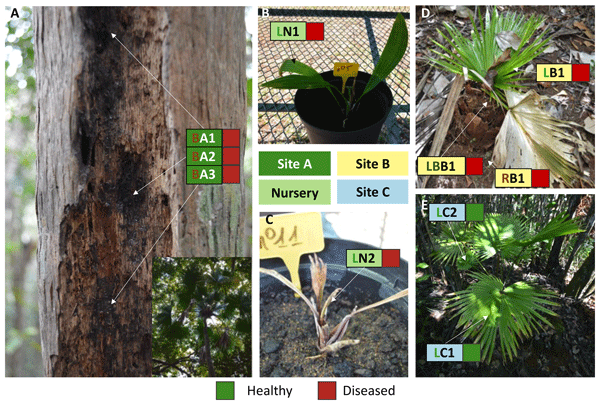
As a result of illegal harvesting and clandestine collection, the geographic coordinates of all sites are withheld. On the adult tree, three samples of bark tissue were collected in order to represent the full extent of the infected area (Fig. 1A). Samples on healthy bark tissues were not taken on other parts of the tronc to avoid any scars and injuries on this unique remaining adult specimen. Pieces of leaves were collected on the two seedlings in nursery (Fig. 1B–C). For the diseased sapling on site B (Fig. 1D), which is the only known individual in this locality (discovered in 2018 and currently dead), samples were collected from different parts: one from a leaf, one from the base of palm and one from aerial roots. The sapling in site C (Fig. 1E) was the only one that was considered healthy. In this specimen, to be as non-invasive as possible, the sampling was restricted to the leaves. All samples were collected in June 2019 and were immediately preserved in 70 % ethanol for further processing DNA extraction and metabarcoding Illumina sequencing. The limited sample size reflects both the extremely small number of surviving individuals and the deliberate use of minimally invasive sampling to avoid causing additional injury or physiological stress to this critically endangered species.
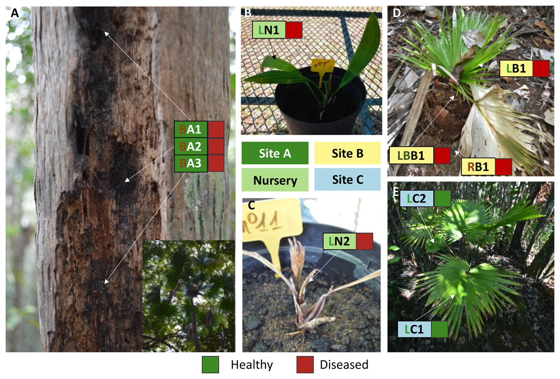
Figure 1(A) Bark tissue exhibiting symptoms of disease for the unique known Saribus jeanneneyi adult individual (site A); (B) diseased seedling and (C) severely infected seedling produced in nursery. (D) Sapling showing leave disease symptoms (site B); (E) sapling harboring no signs of infections (site C); Rectangulars represent the sampling area; the first letter indicates the tissue type collected (B: bark; L: leaf; LB: leaf base; R: roots), and the second letter the site (A: Site A; B: Site B; C: Site C; N: Nursery). Remark: The individual sapling presented in (D) is now recorded as dead, as well as the two nursery seedlings.
2.2 DNA extraction and metabarcoding sequencing
Samples were ground with liquid nitrogen and DNA was thereafter extracted using the Wizard genomic DNA purification kit (Promega) as described in Carriconde et al. (2008). Then, libraries generation and sequencing were realized as previously described in Fernandez Nuñez et al. (2021). Briefly, metabarcoding is a molecular approach that amplifies short, standardized DNA regions (“barcodes”) directly from environmental or host tissue samples to simultaneously identify the many microorganisms present, without the need to culture them individually. For fungi, we targeted the ITS2 region, a widely used fungal barcode (Schoch et al., 2012); for bacteria, we targeted the V4 region of the 16S rRNA gene, the standard bacterial barcode (Caporaso et al., 2011). After sequencing, reads were clustered into Amplicon Sequence Variants (ASVs), each representing a distinct microbial taxon, and each ASV was assigned a taxonomic identity ((phylum, genus and, where possible, species) by comparison against reference databases. Approximately 6 million paired reads of 250 bp length were obtained for both ITS2 (fungi) and V4 (bacteria) in independent sequencing runs.
2.3 Bioinformatics and statistics
All steps were realized as previously described in Stenger et al. (2025a), except that rarefaction was not carried out to obtain all the diversity possible for pathogen research. The bacteria analysis were done with updated database and QIIME version, as explained in Stenger et al. (2025b). This allows us to reconstruct, for each sample, the all communities of fungi and bacteria, the relative abundances of the different observed taxa, as well as to compare these communities between diseased and healthy tissues.
2.4 Co-occurrence analysis
To identify which microbial taxa tend to occur together, and therefore might interact within the diseased bark, we additionally performed a co-occurrence network analysis: taxa whose abundances rose and fell together across samples (based on statistically significant correlations) were linked in a network, allowing us to detect tightly interconnected “hub” taxa that are good candidates for playing a central ecological role in the disease process. Counts were converted to relative abundances by dividing each ASV count by the total reads per sample. To address compositionality and zero inflation, a centered log-ratio (CLR) transformation was applied after adding a small pseudocount (0.01 × minimum nonzero value). CLR-transformed matrices were used for network analysis. For network construction, a combined matrix of bacteria and fungi CLR abundances was created for bark samples only. Pairwise Spearman rank correlations were calculated between all taxa across bark samples. P-values were adjusted by the Benjamini-Hochberg method. Significant correlations were retained where and adjusted p≤0.05. An undirected, weighted network was constructed in igraph: nodes represent ASVs (annotated at the genus level), and edges represent significant correlations. For centrality and pathogen candidate scoring, node centrality metrics (degree, betweenness, closeness, eigenvector centrality) were computed using absolute edge weights. Mean CLR abundances in diseased versus healthy bark were compared to generate an enrichment ratio (log2[diseased healthy + 0.001]). Candidate pathogens were ranked by a composite score combining scaled centrality metrics, positive enrichment, and a bonus for membership in known pathogenic genera (e.g., Pseudomonas, Fusarium, etc.). For community detection and subnetwork extraction, high-degree hub taxa (top 5 % by degree) and their first-order neighbors were extracted to form a pathogen-focused subnetwork. Community structure was visualized using the Fruchterman-Reingold layout with absolute edge weights for repulsion, and node labels were applied only to hub genera to avoid clutter.
3.1 Tissue microbial communities
Before looking for the specific presence of pathogens, we will briefly describe the microbial communities present in the tissues (Fig. 2). Fungal community composition was dominated by three major phyla across all samples (Fig. 3, upper panel). Ascomycota constituted the most abundant phylum, with reads ranging from 7582 in sample LR2 to 44 730 in sample LN2. Diseased bark samples from Site A (BA1, BA2, BA3) contained 19 134–42 042 Ascomycota reads, representing the dominant component in all bark tissues. Basidiomycota formed the second most abundant phylum, with the highest abundance recorded in sample LR2 (57 722 reads) and minimum abundances in bark samples LN1 (215 reads). Diseased bark samples from Site A showed lower Basidiomycota abundances (733–2050 reads). Mucoromycota was detected at minimal levels, with only four reads recorded in sample LR1 and complete absence in all other samples. The sample LR2 exhibited a balanced composition between Ascomycota (7582 reads) and Basidiomycota (57 722 reads), contrasting with the Ascomycota-dominated patterns observed in all other samples.
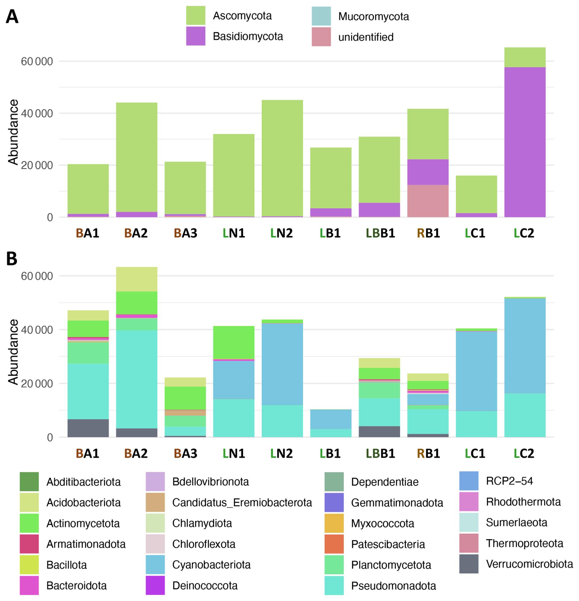
Figure 2Stacked barplots illustrating the absolute abundances of fungal (A) and bacterial (B) phyla in each sample. Labels: first letter is for tissue (B: bark, L: leaf, LB: leaf base,R: roots), the second letter is for the site (A: Site A, B: Site B, C: Site C, N: Nursery).
Bacterial community composition was dominated by three major phyla with distinct distribution patterns (Fig. 3, lower panel). Pseudomonadota constituted the most abundant phylum across most samples, with the highest abundance recorded in sample BA2 (36 454 reads) and substantial presence in diseased bark samples from Site A (3390–36 454 reads). Cyanobacteriota formed the second most abundant phylum, exhibiting the highest abundances in leaf tissues, particularly samples LN2 (30 274 reads), LR1 (29 597 reads), and LR2 (35 276 reads), while remaining at low levels in bark samples (33–260 reads). Actinomycetota represented the third major phylum, with pronounced abundance in bark tissues from diseased Site A samples (6173–8658 reads) and moderate levels in healthy samples LN1 (12 180 reads) and RB1 (3168 reads). The bacterial community structure in diseased bark samples (BA1, BA2, BA3) was characterized by high Pseudomonadota dominance, moderate Actinomycetota representation, and minimal Cyanobacteriota presence, contrasting with the Cyanobacteriota-dominated profiles observed in leaf tissues from other sites.
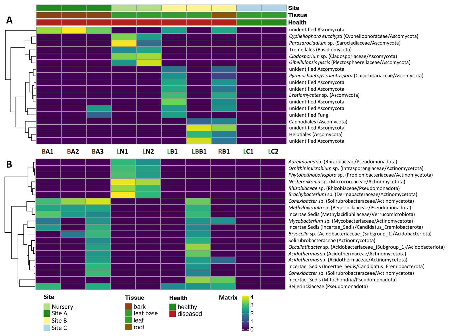
Figure 3Heatmaps representing the CLR-transformed abundance profiles of fungal (A) and bacterial (B) taxa detected across all samples (log2FoldChange > and Padj < 0.05 for diseased vs. healthy). Columns correspond to individual samples, grouped by site (Nursery, Site A, Site B, Site C), tissue type (bark, leaf base, leaf, root), and health status (healthy or diseased), as indicated by the color-coded annotations. Rows show the most abundant or discriminatory taxa, labeled at the genus or higher taxonomic rank, with associated family and phylum indicated in parentheses. Hierarchical clustering was performed independently for each panel using Euclidean distance and complete linkage. Yellow to purple color scale indicates increasing to decreasing CLR abundance, respectively.
CLR-transformed abundance profiles revealed pronounced shifts in both fungal and bacterial communities between healthy and symptomatic trees across sites and tissues. Fungal assemblages of diseased trees were enriched for several Ascomycota lineages, including Cyphellophora eucalypti, Gibellulopsis piscis, Parasarocladium sp., Pyrenochaetopsis leptospora, Leotiomycetes sp. and unknown Ascomycota. Bacterial communities showed a similar pattern, with diseased samples characterized by increased CLR abundance of multiple Actinomycetota and Acidobacteriota genera, such as Acidothermus, Brachybacterium, Bryocella, Conexibacter, Mycobacterium, Nesterenkonia, Occallatibacter and Ornithinimicrobium.
3.2 Co-occurrence analysis
The co-occurrence network revealed a single, highly central cluster of microbial taxa predominantly associated with bark samples from Site A. This cluster alone exhibited sufficient connectivity to meet our top-degree threshold, indicating that these taxa form a tightly interlinked consortium in diseased bark. The remaining clusters (which represent communities from other tissues (leaf, root) or other sites) did not reach the same degree of centrality and therefore were not annotated. The presence of these multiple clusters reflects niche differentiation and site-specific community structure: tightly knit modules correspond to particular ecological niches or health states. In particular, the annotated hub cluster suggests that a limited set of bacterial and fungal taxa co-associate intensively under diseased conditions. These hub taxa formed the central interconnected cluster and included key fungal genera such as Gibellulopsis, Parasarcocladium, Cladosporium, Xenoacremonium, and Mycosphaerella were identified as central nodes. Additionally, bacterial genera including Brachybacterium, Occallatibacter, Jatrophihabitans, Bryocella, Conexibacter, Halomonas, Methylovirgula, Asticcacaulis, Fulvimarina, Phytoactinoporlyspora, Ornithinimicrobium, and Acidothermus were identified as central nodes. These labeled taxa represented 25 % (74 out of 296 nodes) of the total network (Fig. 4) but accounted for the majority of significant correlations, indicating their role as keystone species within the microbial community structure. Peripheral clusters with fewer or less central nodes likely represent background microbiota or taxa whose interactions are more localized to non-bark environments or healthy samples. Consequently, focusing on the central, annotated module provides the most direct path toward identifying the microbial drivers of bark disease in Site A.
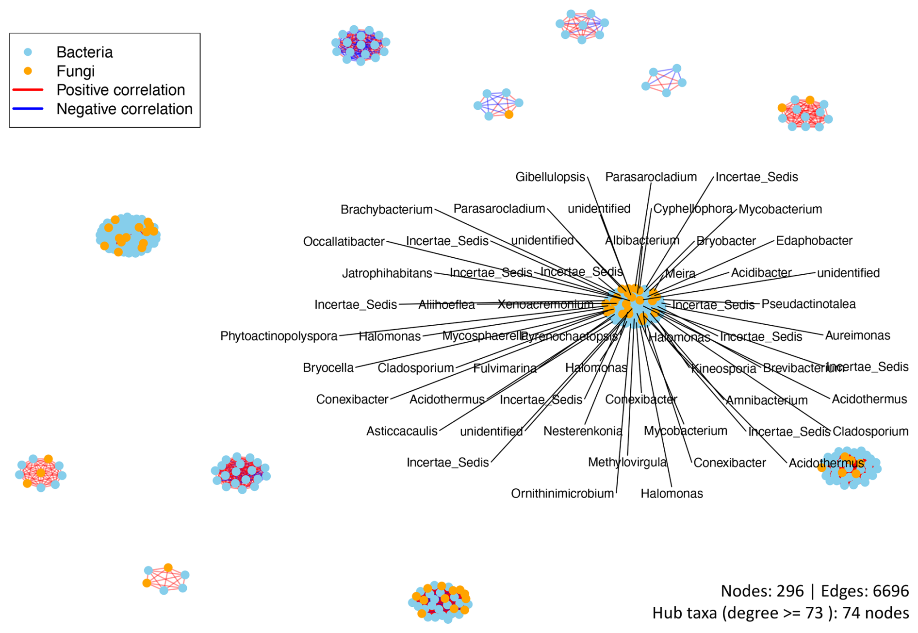
Figure 4Network diagram illustrating significant co-occurrence relationships among bacterial and fungal genera in samples. Nodes are colored by kingdom; the sky-blue nodes represent bacteria and the orange nodes represent fungi. Edges indicate strong, statistically significant Spearman correlations: red lines correspond to positive associations, while blue lines correspond to negative associations. The central hub cluster comprises 74 highly connected taxa (degree ≥ 73), reflecting dominant microbial interactions in the dataset. Peripheral clusters highlight additional community modules. The counts of nodes (296), edges (6696), and hub taxa are included for reference.
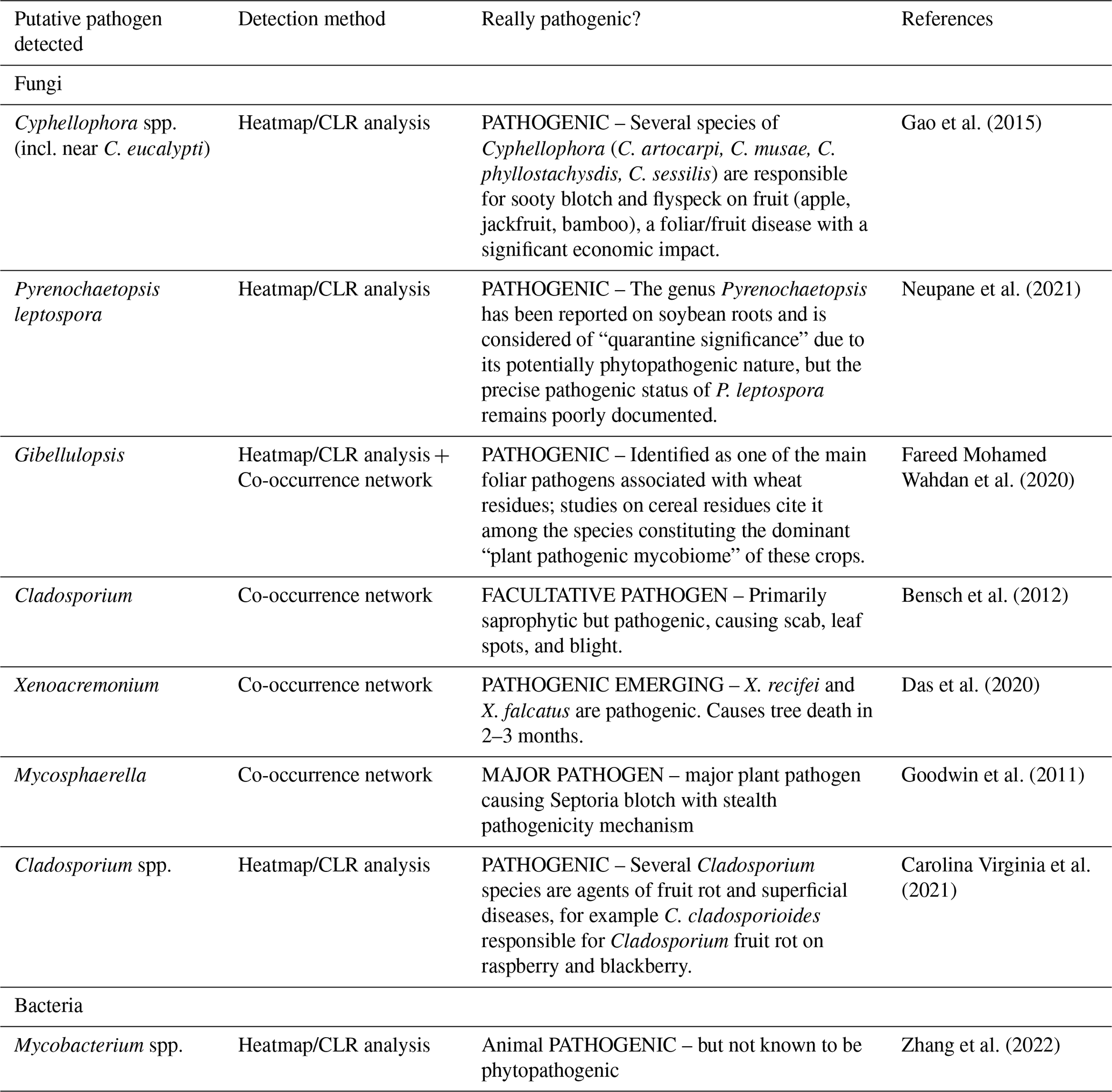
Moreover, heatmap-based CLR profiles and co-occurrence network analysis consistently recovered a small set of fungal genera with documented plant pathogenicity (Table 1), including Cyphellophora, Pyrenochaetopsis, Gibellulopsis, Cladosporium, Xenoacremonium, and Mycosphaerella, as being enriched and/or centrally embedded within the diseased microbiome of S. jeanneneyi.
Table 1Overview of putative pathogens detected in S. jeanneneyi bark and assessment of their plant pathogenic status (Fungi and Bacteria), listing their method of detection, assessment of genuine pathogenic potential, and a representative literature reference.
4.1 Polymicrobial bark disease in the last remining S. jeanneneyi adult palm
Our results indicate that bark disease in the last remaining wild adult S. jeanneneyi is best explained by a polymicrobial syndrome rather than a single causal agent. Diseased harboured strongly structured fungal and bacterial communities, with a small set of Ascomycota genera (Cyphellophora, Gibellulopsis, Pyrenochaetopsis, Cladosporium, Xenoacremonium and Mycosphaerella) simultaneously enriched in CLR-based heatmaps and centrally embedded within the co-occurrence network. All these genera contain well-documented plant fungal pathogens responsible for foliar, fruit and stem diseases in other hosts (e.g. sooty blotch and flyspeck, septoria blotch, fruit rots, rapid tree decline) (Table 1), supporting their interpretation as genuine or emerging phytopathogens in S. jeanneneyi (Bensch et al., 2012; Carolina Virginia et al., 2021; Das et al., 2020; Fareed Mohamed Wahdan et al., 2020; Gao et al., 2015; Goodwin et al., 2011; Neupane et al., 2021). This pattern is consistent with increasing evidence that many plant diseases reflect pathogenic consortia embedded within dysbiotic microbiomes rather than the action of a single invader (Cobo-Díaz et al., 2019; Fareed Mohamed Wahdan et al., 2020; Spragge et al., 2023).
4.2 Contrasting roles of fungi and bacteria
Fungi and bacteria appear, here, to play contrasting ecological roles in the disease-associated microbiome. Indeed, fungal assemblages in diseased bark were dominated by Ascomycota lineages with clear pathogenic credentials in other systems, including Mycosphaerella, a major septoria blotch agent, and Xenoacremonium, an emerging tree pathogen, together with Cyphellophora, Gibellulopsis, Pyrenochaetopsis, and Cladosporium, all implicated in foliar and stem diseases across crops (Bensch et al., 2012; Carolina Virginia et al., 2021; Das et al., 2020; Fareed Mohamed Wahdan et al., 2020; Gao et al., 2015; Goodwin et al., 2011; Neupane et al., 2021). By contrast, the main bacterial hubs were dominated by taxa typically considered saprophytic, oligotrophic or potentially beneficial, such as Acidothermus, Brachybacterium, Bryocella, Jatrophihabitans, Halomonas, Methylovirgula, Occallatibacter, Ornithinimicrobium, suggesting roles as decomposers, commensals or antagonists rather than primary drivers of bark necrosis (Hudson et al., 2023; Maciag et al., 2023). This interpretation aligns with work showing that certain bacterial guilds can protect plants against pathogens through nutrient competition or direct antagonism, and that increased microbiome diversity can buffer hosts against disease (Spragge et al., 2023; West et al., 2019).
At the same time, diseased bark showed a marked depletion of Cyanobacteriota and an enrichment of Actinomycetota and Acidobacteriota, indicating a shift towards a more heterotrophic, stress-adapted microbiome typical of decaying or pathogen-impacted tissues. Such compositional changes are increasingly recognized as signatures of plant microbiome dysbiosis under disease or environmental stress (Arnault et al., 2023; Gao et al., 2021; Yu et al., 2026). In S. jeanneneyi, this suggests that a small suite of fungal pathogens exploits a reorganized microbial landscape, while bacterial associates mainly modulate, rather than initiate, bark decline.
4.3 Conservation and management implications
For a micro-endemic palm represented by a single adult and a few juveniles, the emergence of a fungus-dominated polymicrobial bark disease poses an immediate extinction risk.
Concretely, we recommend three immediate actions for conservation managers overseeing the remaining wild individuals and the ex situ collection. First, symptomatic bark and foliar tissue should be regularly monitored visually and, where feasible, resampled for metabarcoding to track whether the fungal consortium identified here (Cyphellophora, Gibellulopsis, Xenoacremonium, Mycosphaerella) is spreading or evolving, given the very rapid tree decline reported for related Xenoacremonium infections elsewhere (Das et al., 2020). Second, rather than applying broad-spectrum biocides, conservation managers should consider strictly localised and monitored trials of registered antifungal products, guided by isolate-level identification and fungicide-sensitivity testing (Ishii and Hollomon, 2015; Lucas et al., 2015). At present, our metabarcoding data do not justify selecting a fungicide against a particular candidate genus. Third, given the very small number of surviving individuals, any newly produced seedlings or cuttings should be physically isolated from the diseased adult and from each other where possible, to avoid potential horizontal transmission of the candidate fungal pathogens before their causal role is experimentally confirmed. Finally, because fungal identification here relies on correlational, sequence-based evidence rather than pathogenicity trials, we stress that these three fungal genera should be treated as priority targets for confirmatory testing (e.g., isolation and inoculation trials) rather than as definitively proven causal agents, so that management resources are not misallocated. In parallel, managing soils and litter to support diverse saprophytic and endophytic bacteria could contribute indirectly to disease suppression, in line with evidence that microbiome diversity can protect hosts against pathogen invasion (Sallam et al., 2023; Spragge et al., 2023). For a species where every individual is irreplaceable, integrating high-throughput microbiome diagnostics into routine health monitoring and conservation planning appears essential rather than optional (West et al., 2019).
4.4 Outlook
This study illustrates how high-throughput metabarcoding combined with compositional analyses and network inference can rapidly identify candidate pathogens and their microbial context in an endangered plant with extremely limited sample availability, complementing recent calls to use high-throughput sequencing tools into plant disease diagnostics (Cobo-Díaz et al., 2019; Hariharan and Prasannath, 2021). Future work should build on these insights by (i) developing species-specific molecular assays for the main fungal candidates and by testing microbiome-informed management strategies within ongoing ex situ and in situ conservation programmes; (ii) running metatranscriptomics analysis to distinguish metabolically active pathogens from dormant organisms; and (iii) realized cultivation-independent pathogenicity assays using plant tissue cultures to validate disease-causing potential without requiring traditional Koch's postulates (Hariharan and Prasannath, 2021; Sudakov et al., 2024).
All code and scripts are available at https://github.com/PLStenger/Saribus_jeanneneyi (last access: 4 November 2026) and https://doi.org/10.5281/zenodo.23949781 (Stenger, 2026).
Raw sequencing data are deposited in the Sequence Read Archive, the National Center for Biotechnology Information (BioProject: PRJNA1463181). All other data files are available at https://github.com/PLStenger/Saribus_jeanneneyi (last access: 4 November 2026) and https://doi.org/10.5281/zenodo.23949781 (Stenger, 2026).
FC conceived, performed the field sampling and supervised this study. PLS analysed the data, and PLS and FC wrote the manuscript.
The contact author has declared that neither of the authors has any competing interests.
Publisher's note: Copernicus Publications remains neutral with regard to jurisdictional claims made in the text, published maps, institutional affiliations, or any other geographical representation in this paper. The authors bear the ultimate responsibility for providing appropriate place names. Views expressed in the text are those of the authors and do not necessarily reflect the views of the publisher.
We would like to thank Arnaud Lespes from the South Province (Parc Provincial de la Rivière Blue) and Stéphane McCoy from Prony Resources (Département Developpement Durable) from bringing us to the different field sites. We are particularly grateful for the information provided on Saribus jeanneneyi by A. Lespes. We thank Kelly Dink (IAC) for extracting the DNA at the Platform du Vivant (PFVNC, Nouméa, New Caledonia) under the supervision of FC. We also gratefully thank Pierre Mournet of the Regional Genotyping Platform (GPTR Génotypage, https://umr-agap.cirad.fr/en/platforms/regional-platforms/genotyping, last access: 4 November 2026) of the UMR AGAP (CIRAD-INRAE-Montpellier SupAgro) for performing the libraries generation and sequencing. We also thank Jérôme Lefevre for allowing us to run the analysis on the Nouméa IRD's cluster.
This paper was edited by Roland Brandl and reviewed by Roland Brandl and Jutta Stadler.
Arnault, G., Mony, C., and Vandenkoornhuyse, P.: Plant microbiota dysbiosis and the Anna Karenina Principle, Trends Plant Sci., 28, 18–30, https://doi.org/10.1016/j.tplants.2022.08.012, 2023.
Bensch, K., Braun, U., Groenewald, J. Z., and Crous, P. W.: The genus Cladosporium, Stud. Mycol., 72, 1–401, https://doi.org/10.3114/sim0003, 2012.
Bour, A.: Saribus jeanneneyi at the Botanical Garden of Nancy, Palms, 58, 1–4, 2014.
Caporaso, J. G., Lauber, C. L., Walters, W. A., Berg-Lyons, D., Lozupone, C. A., Turnbaugh, P. J., Fierer, N., and Knight, R.: Global patterns of 16S rRNA diversity at a depth of millions of sequences per sample, P. Natl. Acad. Sci. USA, 108, 4516–4522, https://doi.org/10.1073/pnas.1000080107, 2011.
Carolina Virginia, T., Javier Néstor, A., Adrián Dario, C., and Graciela Noemí, P.: Cladosporium species causing “Cladosporium rot” on “Bosc” pear fruit in Argentina, Rev. Argent. Microbiol., 53, 75–77, https://doi.org/10.1016/j.ram.2019.11.006, 2021.
Carriconde, F., Gardes, M., Jargeat, P., Heilmann-Clausen, J., Mouhamadou, B., and Gryta, H.: Population evidence of cryptic species and geographical structure in the cosmopolitan ectomycorrhizal fungus, Tricholoma scalpturatum, Microb. Ecol., 56, 513–524, https://doi.org/10.1007/s00248-008-9370-2, 2008.
Cobo-Díaz, J. F., Baroncelli, R., Le Floch, G., and Picot, A.: Combined Metabarcoding and Co-occurrence Network Analysis to Profile the Bacterial, Fungal and Fusarium Communities and Their Interactions in Maize Stalks, Front. Microbiol., 10, 261, https://doi.org/10.3389/fmicb.2019.00261, 2019.
Das, K., Lee, S. Y., and Jung, H. Y.: Molecular and Morphological Characterization of Two Novel Species Collected from Soil in Korea, Mycobiology, 48, 9–19, https://doi.org/10.1080/12298093.2019.1695717, 2020.
Fareed Mohamed Wahdan, S., Hossen, S., Tanunchai, B., Schädler, M., Buscot, F., and Purahong, W.: Future Climate Significantly Alters Fungal Plant Pathogen Dynamics during the Early Phase of Wheat Litter Decomposition, Microorganisms, 8, https://doi.org/10.3390/microorganisms8060908, 2020.
Fernandez Nuñez, N., Maggia, L., Stenger, P.-L., Lelievre, M., Letellier, K., Gigante, S., Manez, A., Mournet, P., Ripoll, J., and Carriconde, F.: Potential of high-throughput eDNA sequencing of soil fungi and bacteria for monitoring ecological restoration in ultramafic substrates: The case study of the New Caledonian biodiversity hotspot, Ecol. Eng., 173, https://doi.org/10.1016/j.ecoleng.2021.106416, 2021.
Gao, L., Ma, Y., Zhao, W., Wei, Z., Gleason, M. L., Chen, H., Hao, L., Sun, G., and Zhang, R.: Three New Species of Cyphellophora (Chaetothyriales) Associated with Sooty Blotch and Flyspeck, PLoS One, 10, e0136857, https://doi.org/10.1371/journal.pone.0136857, 2015.
Gao, M., Xiong, C., Gao, C., Tsui, C. K. M., Wang, M.-M., Zhou, X., Zhang, A.-M., and Cai, L.: Disease-induced changes in plant microbiome assembly and functional adaptation, Microbiome, 9, 187, https://doi.org/10.1186/s40168-021-01138-2, 2021.
Goodwin, S. B., M'Barek, S. Ben, Dhillon, B., Wittenberg, A. H. J., Crane, C. F., Hane, J. K., Foster, A. J., van der Lee, T. A. J., Grimwood, J., Aerts, A., Antoniw, J., Bailey, A., Bluhm, B., Bowler, J., Bristow, J., van der Burgt, A., Canto-Canché, B., Churchill, A. C. L., Conde-Ferràez, L., Cools, H. J., Coutinho, P. M., Csukai, M., Dehal, P., de Wit, P., Donzelli, B., van de Geest, H. C., van Ham, R. C. H. J., Hammond-Kosack, K. E., Henrissat, B., Kilian, A., Kobayashi, A. K., Koopmann, E., Kourmpetis, Y., Kuzniar, A., Lindquist, E., Lombard, V., Maliepaard, C., Martins, N., Mehrabi, R., Nap, J. P. H., Ponomarenko, A., Rudd, J. J., Salamov, A., Schmutz, J., Schouten, H. J., Shapiro, H., Stergiopoulos, I., Torriani, S. F. F., Tu, H., de Vries, R. P., Waalwijk, C., Ware, S. B., Wiebenga, A., Zwiers, L. H., Oliver, R. P., Grigoriev, I. V., and Kema, G. H. J.: Finished genome of the fungal wheat pathogen Mycosphaerella graminicola reveals dispensome structure, chromosome plasticity, and stealth pathogenesis, PLoS Genet., 7, https://doi.org/10.1371/journal.pgen.1002070, 2011.
Hariharan, G. and Prasannath, K.: Recent Advances in Molecular Diagnostics of Fungal Plant Pathogens: A Mini Review, Front. Cell. Infect. Microbiol., 10, 1–14, https://doi.org/10.3389/fcimb.2020.600234, 2021.
Henry, B.: New Caledonia, a Jewel of Palm, Palms, 65, 132–146, 2021.
Hudson, J. E., Levia, D. F., Yoshimura, K. M., Gottel, N. R., Hudson, S. A., and Biddle, J. F.: Mapping bark bacteria: initial insights of stemflow-induced changes in bark surface phyla, Microbiol. Spectr., 11, e0356223, https://doi.org/10.1128/spectrum.03562-23, 2023.
Ishii, H. and Hollomon, D. W.: Fungicide resistance in plant pathogens: principles and a guide to practical management, Springer, ISBN 978-4-431-55642-8, https://doi.org/10.1007/978-4-431-55642-8, 2015.
Ititiaty, Y., Gâteblé, G., Fogliani, B., Mccoy, S., Manaute, J., and Lespes, A.: Germination, cultivation and ex situ conservation of an extremely rare palm endemic to New Caledonia, in: 11th International Conference on Serpentine Ecology (ICSE2025), 1, https://hal.science/hal-05508322 (last access: 4 November 2026), 2025.
Jiang, J., Hu, X., Ji, X., and Chen, H.: High throughput sequencing technology facility research of genomic modification crop cultivation influencing soil microbe, Front. Plant Sci., 14, 1208111, https://doi.org/10.3389/fpls.2023.1208111, 2023.
Lucas, J. A., Hawkins, N. J., and Fraaije, B. A.: Chapter Two – The Evolution of Fungicide Resistance, in: Advances in applied microbiology, vol. 90, edited by: Sariaslani, S. and Gadd, G. M. B. T.-A., Academic Press, 29–92, https://doi.org/10.1016/bs.aambs.2014.09.001, 2015.
Maciag, T., Kozieł, E., Rusin, P., Otulak-Kozieł, K., Jafra, S., and Czajkowski, R.: Microbial Consortia for Plant Protection against Diseases: More than the Sum of Its Parts., Int. J. Mol. Sci., 24, https://doi.org/10.3390/ijms241512227, 2023.
Nafea, A. M., Wang, Y., Wang, D., Salama, A. M., Aziz, M. A., Xu, S., and Tong, Y.: Application of next-generation sequencing to identify different pathogens, Front. Microbiol., 14, 1329330, https://doi.org/10.3389/fmicb.2023.1329330, 2023.
Neupane, A., Bulbul, I., Wang, Z., Lehman, R. M., Nafziger, E., and Marzano, S. Y. L.: Long term crop rotation effect on subsequent soybean yield explained by soil and root-associated microbiomes and soil health indicators, Sci. Rep., 11, 1–13, https://doi.org/10.1038/s41598-021-88784-6, 2021.
Pintaud, J.-C., Jaffré, T., and Veillon, J.-M.: Conservation status of New Caledonia palms, Pacific Conserv. Biol., 5, 9–15, 1999.
Sallam, N. M. A., AbdElfatah, H.-A. S., Khalil Bagy, H. M. M., Elfarash, A., Abo-Elyousr, K. A. M., Sikora, E. J., and Sallam, A.: Exploring the mechanisms of endophytic bacteria for suppressing early blight disease in tomato (Solanum lycopersicum L.), Front. Microbiol., 14, 1184343, https://doi.org/10.3389/fmicb.2023.1184343, 2023.
Schoch, C. L., Seifert, K. A., Huhndorf, S., Robert, V., Spouge, J. L., Levesque, C. A., Chen, W., Fungal Barcoding Consortium, Bolchacova, E., Voigt, K., Crous, P. W., Miller, A. N., Wingfield, M. J., Aime, M. C., An, K.-D., Bai, F.-Y., Barreto, R. W., Begerow, D., Bergeron, M.-J., Blackwell, M., Boekhout, T., Bogale, M., Boonyuen, N., Burgaz, A. R., Buyck, B., Cai, L., Cai, Q., Cardinali, G., Chaverri, P., Coppins, B. J., Crespo, A., Cubas, P., Cummings, C., Damm, U., de Beer, Z. W., de Hoog, G. S., Del-Prado, R., Dentinger, B., Diéguez-Uribeondo, J., Divakar, P. K., Douglas, B., Dueñas, M., Duong, T. A., Eberhardt, U., Edwards, J. E., Elshahed, M. S., Fliegerova, K., Furtado, M., García, M. A., Ge, Z.-W., Griffith, G. W., Griffiths, K., Groenewald, J. Z., Groenewald, M., Grube, M., Gryzenhout, M., Guo, L.-D., Hagen, F., Hambleton, S., Hamelin, R. C., Hansen, K., Harrold, P., Heller, G., Herrera, C., Hirayama, K., Hirooka, Y., Ho, H.-M., Hoffmann, K., Hofstetter, V., Högnabba, F., Hollingsworth, P. M., Hong, S.-B., Hosaka, K., Houbraken, J., Hughes, K., Huhtinen, S., Hyde, K. D., James, T., Johnson, E. M., Johnson, J. E., Johnston, P. R., Jones, E. B. G., Kelly, L. J., Kirk, P. M., Knapp, D. G., Kõljalg, U., Kovács, G. M., Kurtzman, C. P., Landvik, S., Leavitt, S. D., Liggenstoffer, A. S., Liimatainen, K., Lombard, L., Luangsa-ard, J. J., Lumbsch, H. T., Maganti, H., Maharachchikumbura, S. S. N., Martin, M. P., May, T. W., McTaggart, A. R., Methven, A. S., Meyer, W., Moncalvo, J.-M., Mongkolsamrit, S., Nagy, L. G., Nilsson, R. H., Niskanen, T., Nyilasi, I., Okada, G., Okane, I., Olariaga, I., Otte, J., Papp, T., Park, D., Petkovits, T., Pino-Bodas, R., Quaedvlieg, W., Raja, H. A., Redecker, D., Rintoul, T. L., Ruibal, C., Sarmiento-Ramírez, J. M., Schmitt, I., Schüßler, A., Shearer, C., Sotome, K., Stefani, F. O. P., Stenroos, S., Stielow, B., Stockinger, H., Suetrong, S., Suh, S.-O., Sung, G.-H., Suzuki, M., Tanaka, K., Tedersoo, L., Telleria, M. T., Tretter, E., Untereiner, W. A., Urbina, H., Vágvölgyi, C., Vialle, A., Vu, T. D., Walther, G., Wang, Q.-M., Wang, Y., Weir, B. S., Weiß, M., White, M. M., Xu, J., Yahr, R., Yang, Z. L., Yurkov, A., Zamora, J.-C., Zhang, N., Zhuang, W.-Y., and Schindel, D.: Nuclear ribosomal internal transcribed spacer (ITS) region as a universal DNA barcode marker for Fungi, P. Natl. Acad. Sci. USA, 109, 6241–6246, https://doi.org/10.1073/pnas.1117018109, 2012.
Spragge, F., Bakkeren, E., Jahn, M. T., Araujo, E. B. N., Pearson, C. F., Wang, X., Pankhurst, L., Cunrath, O., and Foster, K. R.: Microbiome diversity protects against pathogens by nutrient blocking, Science, 382, https://doi.org/10.1126/science.adj3502, 2023.
Stenger, P.-L.: PLStenger/Saribus_jeanneneyi: DOI Web Ecology (Version DOI_Web_Ecology), Zenodo [data set] and [code], https://doi.org/10.5281/zenodo.23949781, 2026.
Stenger, P.-L., Léopold, A., Dinh, K., Mournet, P., Robert, N., Drouin, J., Wamejonengo, J., Russet, S., Ibanez, T., Maggia, L., and Carriconde, F.: Advancing biomonitoring of eDNA studies with the Anaconda R package: integrating soil and One Health perspectives in the face of evolving traditional agriculture practices, PLoS One, 20, e0311986, https://doi.org/10.1371/journal.pone.0311986, 2025a.
Stenger, P.-L., Bonneville, C. D., Anton-Leberre, V., Cleguer, C., Fauvelot, C., Garrigue, C., Derville, S., and Majorel, C.: Exploring the skin Microbiome of free-ranging Dugong dugon in new Caledonia, Mar. Biol., 172, 1–23, https://doi.org/10.1007/s00227-025-04696-3, 2025b.
Sudakov, K., Yasuor, H., Jaiswal, A. K., Mordukhovich, G., Buchshtab, O., Minz, D., and Frenkel, O.: Incorporating Culture-Independent Methods to Identify Net Syndrome-Associated Fusarium Species in Peanuts, Phytobiomes J., 8, 318–326, https://doi.org/10.1094/PBIOMES-10-23-0111-R, 2024.
West, A. G., Waite, D. W., Deines, P., Bourne, D. G., Digby, A., McKenzie, V. J., and Taylor, M. W.: The microbiome in threatened species conservation, Biol. Conserv., 229, 85–98, https://doi.org/10.1016/j.biocon.2018.11.016, 2019.
Yu, C., Wang, M., Zhao, M., Zhang, S., Cao, M., Liu, Z., Jiang, J., Zhang, Y., Pan, Y., and Zhao, X.: Root-associated bacterial community dynamics and assembly mechanisms in healthy and root rot-infected soybeans, Front. Microbiol., 17, 1789440, https://doi.org/10.3389/fmicb.2026.1789440, 2026.
Zhang, H., Liu, M., Fan, W., Sun, S., and Fan, X.: The impact of Mycobacterium tuberculosis complex in the environment on one health approach, Front. Public Heal., 10, https://doi.org/10.3389/fpubh.2022.994745, 2022.